[English] 日本語
Yorodumi
- PDB-3eyy: Structural basis for the specialization of Nur, a nickel-specific... -

+
Open data


ID or keywords:

Loading...

-
Basic information

Entry
Database: PDB / ID: 3eyy
Title
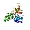
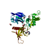
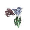
Structural basis for the specialization of Nur, a nickel-specific Fur homologue, in metal sensing and DNA recognition
ComponentsPutative iron uptake regulatory protein
KeywordsTRANSPORT / Nur / nickel-uptake regulator / D-domain / dimerization domain / DB-domain / DNA-binding domain
Function / homology
Function and homology information


regulation of secondary metabolite biosynthetic process / transcription cis-regulatory region binding / DNA-binding transcription factor activity / negative regulation of DNA-templated transcription / DNA binding / zinc ion binding
Similarity search - Function
Ferric-uptake regulator, C-terminal dimerisarion domain / Ferric-uptake regulator, C-terminal domain / Ferric-uptake regulator / Ferric uptake regulator family / Dna Ligase; domain 1 / Winged helix-like DNA-binding domain superfamily/Winged helix DNA-binding domain / Arc Repressor Mutant, subunit A / Winged helix DNA-binding domain superfamily / Winged helix-like DNA-binding domain superfamily / 2-Layer Sandwich ...Ferric-uptake regulator, C-terminal dimerisarion domain / Ferric-uptake regulator, C-terminal domain / Ferric-uptake regulator / Ferric uptake regulator family / Dna Ligase; domain 1 / Winged helix-like DNA-binding domain superfamily/Winged helix DNA-binding domain / Arc Repressor Mutant, subunit A / Winged helix DNA-binding domain superfamily / Winged helix-like DNA-binding domain superfamily / 2-Layer Sandwich / Orthogonal Bundle / Mainly Alpha / Alpha Beta
Similarity search - Domain/homology
MALONATE ION / NICKEL (II) ION / Putative iron uptake regulatory protein
Similarity search - Component
Biological speciesStreptomyces coelicolor (bacteria)
MethodX-RAY DIFFRACTION / SYNCHROTRON / MAD / Resolution: 2.4 Å
AuthorsCha, S.-S. / An, Y.J.
CitationJournal: Nucleic Acids Res. / Year: 2009
Title: Structural basis for the specialization of Nur, a nickel-specific Fur homolog, in metal sensing and DNA recognition
Authors: An, Y.J. / Ahn, B.-E. / Han, A.-R. / Kim, H.-M. / Chung, K.M. / Shin, J.-H. / Cho, Y.-B. / Roe, J.-H. / Cha, S.-S.
History
DepositionOct 22, 2008Deposition site: RCSB / Processing site: PDBJ
Revision 1.0Jun 9, 2009Provider: repository / Type: Initial release
Revision 1.1Jul 13, 2011Group: Version format compliance
Revision 1.2Dec 27, 2023Group: Data collection / Database references / Derived calculations
Category: chem_comp_atom / chem_comp_bond ...chem_comp_atom / chem_comp_bond / database_2 / pdbx_struct_conn_angle / struct_conn / struct_site
Item: _database_2.pdbx_DOI / _database_2.pdbx_database_accession ..._database_2.pdbx_DOI / _database_2.pdbx_database_accession / _pdbx_struct_conn_angle.ptnr1_auth_comp_id / _pdbx_struct_conn_angle.ptnr1_auth_seq_id / _pdbx_struct_conn_angle.ptnr1_label_asym_id / _pdbx_struct_conn_angle.ptnr1_label_atom_id / _pdbx_struct_conn_angle.ptnr1_label_comp_id / _pdbx_struct_conn_angle.ptnr1_label_seq_id / _pdbx_struct_conn_angle.ptnr3_auth_comp_id / _pdbx_struct_conn_angle.ptnr3_auth_seq_id / _pdbx_struct_conn_angle.ptnr3_label_asym_id / _pdbx_struct_conn_angle.ptnr3_label_atom_id / _pdbx_struct_conn_angle.ptnr3_label_comp_id / _pdbx_struct_conn_angle.ptnr3_label_seq_id / _pdbx_struct_conn_angle.value / _struct_conn.pdbx_dist_value / _struct_conn.ptnr1_auth_asym_id / _struct_conn.ptnr1_auth_comp_id / _struct_conn.ptnr1_auth_seq_id / _struct_conn.ptnr1_label_asym_id / _struct_conn.ptnr1_label_atom_id / _struct_conn.ptnr1_label_comp_id / _struct_conn.ptnr1_label_seq_id / _struct_conn.ptnr2_auth_asym_id / _struct_conn.ptnr2_auth_comp_id / _struct_conn.ptnr2_auth_seq_id / _struct_conn.ptnr2_label_asym_id / _struct_conn.ptnr2_label_atom_id / _struct_conn.ptnr2_label_comp_id / _struct_site.pdbx_auth_asym_id / _struct_site.pdbx_auth_comp_id / _struct_site.pdbx_auth_seq_id

-
Structure visualization

Structure viewerMolecule:
MolmilJmol/JSmol

Downloads & links

-
Assembly

Deposited unit
A: Putative iron uptake regulatory protein
B: Putative iron uptake regulatory protein
hetero molecules


Theoretical massNumber of molelcules
Total (without water)33,48818
Polymers32,5092
Non-polymers98016
Water1,47782
1


  • Idetical with deposited unit
  • defined by author&software
TypeNameSymmetry operationNumber
identity operation1_555x,y,z1
Buried area2490 Å2
ΔGint-28 kcal/mol
Surface area14020 Å2
MethodPISA
Unit cell
Length a, b, c (Å)79.164, 79.164, 49.732
Angle α, β, γ (deg.)90.00, 90.00, 120.00
Int Tables number144
Space group name H-MP31

-
Components

-
Protein , 1 types, 2 molecules AB

#1: Protein Putative iron uptake regulatory protein


Mass: 16254.288 Da / Num. of mol.: 2
Source method: isolated from a genetically manipulated source
Source: (gene. exp.) Streptomyces coelicolor (bacteria) / Plasmid: pET-3a / Production host: Escherichia coli (E. coli) / Strain (production host): BL21(DE3) / References: UniProt: Q9K4F8

-
Non-polymers , 6 types, 98 molecules

#2: Chemical ChemComp-NI / NICKEL (II) ION / Nickel


Mass: 58.693 Da / Num. of mol.: 2 / Source method: obtained synthetically / Formula: Ni
#3: Chemical ChemComp-ZN / ZINC ION


Mass: 65.409 Da / Num. of mol.: 2 / Source method: obtained synthetically / Formula: Zn
#4: Chemical
ChemComp-CL / CHLORIDE ION / Chloride


Mass: 35.453 Da / Num. of mol.: 5 / Source method: obtained synthetically / Formula: Cl
#5: Chemical
ChemComp-EDO / 1,2-ETHANEDIOL / ETHYLENE GLYCOL / Ethylene glycol


Mass: 62.068 Da / Num. of mol.: 4 / Source method: obtained synthetically / Formula: C2H6O2
#6: Chemical ChemComp-MLI / MALONATE ION / Malonic acid


Mass: 102.046 Da / Num. of mol.: 3 / Source method: obtained synthetically / Formula: C3H2O4
#7: Water ChemComp-HOH / water / Water


Mass: 18.015 Da / Num. of mol.: 82 / Source method: isolated from a natural source / Formula: H2O

-
Experimental details

-
Experiment

ExperimentMethod: X-RAY DIFFRACTION / Number of used crystals: 1

-
Sample preparation

CrystalDensity Matthews: 2.76 Å3/Da / Density % sol: 55.48 %
Crystal growTemperature: 296.15 K / Method: microbatch / pH: 7
Details: 14% PEG 3350, 0.4M sodium malonate (pH 7), 0.1mM benzamidine hydrochloride, 0.1mM NiCl2, pH 7.0, Microbatch, temperature 296.15K

-
Data collection

DiffractionMean temperature: 100 K
Diffraction sourceSource: SYNCHROTRON / Site: Photon Factory / Beamline: BL-5A / Wavelength: 1.4847, 1.4857, 1.4510
DetectorType: ADSC QUANTUM 210 / Detector: CCD / Date: Oct 6, 2007
RadiationMonochromator: Ni MIRROR / Protocol: MAD / Monochromatic (M) / Laue (L): M / Scattering type: x-ray
Radiation wavelength
IDWavelength (Å)Relative weight
11.48471
21.48571
31.4511
ReflectionResolution: 2.4→50 Å / Num. obs: 18205 / % possible obs: 99.8 % / Observed criterion σ(F): 1 / Observed criterion σ(I): 1 / Biso Wilson estimate: 27.5 Å2
Reflection shellHighest resolution: 2.4 Å / % possible all: 99.8

-
Processing

Software
NameVersionClassification
ADSCQuantumdata collection
SOLVEphasing
CNS1.1refinement
HKL-2000data reduction
HKL-2000data scaling
RefinementMethod to determine structure: MAD / Resolution: 2.4→19.79 Å / Rfactor Rfree error: 0.01 / Data cutoff high absF: 1031976.85 / Data cutoff low absF: 0 / Isotropic thermal model: RESTRAINED / Cross valid method: THROUGHOUT / σ(F): 1.1 / Stereochemistry target values: Engh & Huber
RfactorNum. reflection% reflectionSelection details
Rfree0.264 655 5 %RANDOM
Rwork0.218 ---
obs0.218 13008 95.8 %-
all-13583 --
Solvent computationSolvent model: FLAT MODEL / Bsol: 40.0547 Å2 / ksol: 0.310493 e/Å3
Displacement parametersBiso mean: 55.6 Å2
Baniso -1Baniso -2Baniso -3
1-2.46 Å22.59 Å20 Å2
2--2.46 Å20 Å2
3----4.91 Å2
Refine analyze
FreeObs
Luzzati coordinate error0.37 Å0.31 Å
Luzzati d res low-5 Å
Luzzati sigma a0.37 Å0.32 Å
Refinement stepCycle: LAST / Resolution: 2.4→19.79 Å
ProteinNucleic acidLigandSolventTotal
Num. atoms1960 0 46 82 2088
Refine LS restraints
Refine-IDTypeDev ideal
X-RAY DIFFRACTIONc_bond_d0.007
X-RAY DIFFRACTIONc_angle_deg1.3
X-RAY DIFFRACTIONc_dihedral_angle_d22.9
X-RAY DIFFRACTIONc_improper_angle_d0.74
LS refinement shellResolution: 2.4→2.55 Å / Rfactor Rfree error: 0.034 / Total num. of bins used: 6
RfactorNum. reflection% reflection
Rfree0.341 102 5.1 %
Rwork0.28 1904 -
obs--89.2 %
Xplor file
Refine-IDSerial noParam fileTopol file
X-RAY DIFFRACTION1protein_rep.paramprotein.top
X-RAY DIFFRACTION2ion.paramion.top
X-RAY DIFFRACTION3water_rep.paramwater_rep.top
X-RAY DIFFRACTION4mal.parammal.top
X-RAY DIFFRACTION5edo.paramedo.top

+
About Yorodumi

-
News

-
Feb 9, 2022. New format data for meta-information of EMDB entries

New format data for meta-information of EMDB entries

  • Version 3 of the EMDB header file is now the official format.
  • The previous official version 1.9 will be removed from the archive.

Related info.:EMDB header

External links:wwPDB to switch to version 3 of the EMDB data model

-
Aug 12, 2020. Covid-19 info

Covid-19 info

URL: https://pdbj.org/emnavi/covid19.php

New page: Covid-19 featured information page in EM Navigator.

Related info.:Covid-19 info / Mar 5, 2020. Novel coronavirus structure data

+
Mar 5, 2020. Novel coronavirus structure data

Novel coronavirus structure data

Related info.:Yorodumi Speices / Aug 12, 2020. Covid-19 info

External links:COVID-19 featured content - PDBj / Molecule of the Month (242):Coronavirus Proteases

+
Jan 31, 2019. EMDB accession codes are about to change! (news from PDBe EMDB page)

EMDB accession codes are about to change! (news from PDBe EMDB page)

  • The allocation of 4 digits for EMDB accession codes will soon come to an end. Whilst these codes will remain in use, new EMDB accession codes will include an additional digit and will expand incrementally as the available range of codes is exhausted. The current 4-digit format prefixed with “EMD-” (i.e. EMD-XXXX) will advance to a 5-digit format (i.e. EMD-XXXXX), and so on. It is currently estimated that the 4-digit codes will be depleted around Spring 2019, at which point the 5-digit format will come into force.
  • The EM Navigator/Yorodumi systems omit the EMD- prefix.

Related info.:Q: What is EMD? / ID/Accession-code notation in Yorodumi/EM Navigator

External links:EMDB Accession Codes are Changing Soon! / Contact to PDBj

+
Jul 12, 2017. Major update of PDB

Major update of PDB

  • wwPDB released updated PDB data conforming to the new PDBx/mmCIF dictionary.
  • This is a major update changing the version number from 4 to 5, and with Remediation, in which all the entries are updated.
  • In this update, many items about electron microscopy experimental information are reorganized (e.g. em_software).
  • Now, EM Navigator and Yorodumi are based on the updated data.

External links:wwPDB Remediation / Enriched Model Files Conforming to OneDep Data Standards Now Available in the PDB FTP Archive

-
Yorodumi

Thousand views of thousand structures

  • Yorodumi is a browser for structure data from EMDB, PDB, SASBDB, etc.
  • This page is also the successor to EM Navigator detail page, and also detail information page/front-end page for Omokage search.
  • The word "yorodu" (or yorozu) is an old Japanese word meaning "ten thousand". "mi" (miru) is to see.

Related info.:EMDB / PDB / SASBDB / Comparison of 3 databanks / Yorodumi Search / Aug 31, 2016. New EM Navigator & Yorodumi / Yorodumi Papers / Jmol/JSmol / Function and homology information / Changes in new EM Navigator and Yorodumi

Read more