 Movie
Movie Controller

[English] 日本語
 Yorodumi
- PDB-3eyb: Structural and functional insights into the ligand binding domain... -
+ Open data

- Basic information
| Entry | Database: PDB / ID: 3eyb | ||||||
|---|---|---|---|---|---|---|---|
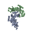
| Title | Structural and functional insights into the ligand binding domain of a non-duplicated RXR from the invertebrate chordate amphioxus | ||||||
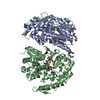
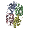
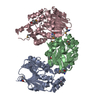
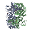
 Components Components | Nuclear hormone receptor RXR | ||||||
 Keywords Keywords | TRANSCRIPTION / Amphioxus / Retinoid X receptor / nuclear receptor / ligand binding / DNA-binding / Metal-binding / Nucleus / Receptor / Transcription regulation / Zinc-finger / Structural Genomics / Structural Proteomics in Europe / SPINE | ||||||
| Function / homology |  Function and homology information Function and homology informationnuclear steroid receptor activity / sequence-specific DNA binding / zinc ion binding / nucleus Similarity search - Function | ||||||
| Biological species |  | ||||||
| Method |  X-RAY DIFFRACTION / SYNCHROTRON / MOLECULAR REPLACEMENT / Resolution: 2.79 Å X-RAY DIFFRACTION / SYNCHROTRON / MOLECULAR REPLACEMENT / Resolution: 2.79 Å | ||||||
 Authors Authors | Tocchini-Valentini, G.D. / Rochel, N. / Moras, D. / Structural Proteomics in Europe (SPINE) | ||||||
 Citation Citation | Journal: J.Biol.Chem. / Year: 2009 Title: Structural and functional insights into the ligand-binding domain of a nonduplicated retinoid X nuclear receptor from the invertebrate chordate amphioxus Authors: Tocchini-Valentini, G.D. / Rochel, N. / Escriva, H. / Germain, P. / Peluso-Iltis, C. / Paris, M. / Sanglier-Cianferani, S. / Van Dorsselaer, A. / Moras, D. / Laudet, V. | ||||||
| History |
|
- Structure visualization
| Structure viewer | Molecule: MolmilJmol/JSmol |
|---|
- Downloads & links
-Download
| PDBx/mmCIF format | 3eyb.cif.gz | 158 KB | Display | PDBx/mmCIF format |
|---|---|---|---|---|
| PDB format | pdb3eyb.ent.gz | 125.7 KB | Display | PDB format |
| PDBx/mmJSON format | 3eyb.json.gz | Tree view | PDBx/mmJSON format | |
| Others |  Other downloads Other downloads |
-Validation report
| Arichive directory | https://data.pdbj.org/pub/pdb/validation_reports/ey/3eybftp://data.pdbj.org/pub/pdb/validation_reports/ey/3eyb | HTTPS FTP |
|---|
-Related structure data
| Related structure data |  1lbd S: Starting model for refinement |
|---|---|
| Similar structure data |


-Links
 PDBj
PDBj
- Assembly
| Deposited unit | 
| ||||||||
|---|---|---|---|---|---|---|---|---|---|
| 1 |
| ||||||||
| Unit cell |
|
-Components
| #1: Protein | Mass: 24697.746 Da / Num. of mol.: 4 / Fragment: UNP residues 266-484, Ligand binding domain Source method: isolated from a genetically manipulated source Source: (gene. exp.) Plasmid: pET15 / Production host:  #2: Water | ChemComp-HOH / |  Mass: 18.015 Da / Num. of mol.: 49 / Source method: isolated from a natural source / Formula: H2O Mass: 18.015 Da / Num. of mol.: 49 / Source method: isolated from a natural source / Formula: H2O |
|---|
-Experimental details
-Experiment
| Experiment | Method: X-RAY DIFFRACTION / Number of used crystals: 1 |
|---|
- Sample preparation
| Crystal | Density Matthews: 2.52 Å3/Da / Density % sol: 51.09 % |
|---|---|
| Crystal grow | Temperature: 290 K / Method: vapor diffusion, hanging drop / pH: 6.5 Details: 1.5 M Na acetate, 0.1 M ADA pH=6.5, VAPOR DIFFUSION, HANGING DROP, temperature 290K |
-Data collection
| Diffraction | Mean temperature: 200 K |
|---|---|
| Diffraction source | Source: SYNCHROTRON / Site: ESRF  / Beamline: ID29 / Beamline: ID29 |
| Detector | Detector: CCD / Date: May 1, 2004 |
| Radiation | Monochromator: Si 111 CHANNEL / Protocol: SINGLE WAVELENGTH / Monochromatic (M) / Laue (L): M / Scattering type: x-ray |
| Radiation wavelength | Relative weight: 1 |
| Reflection | Resolution: 2.79→15 Å / Num. all: 22831 / Num. obs: 22831 / % possible obs: 90.34 % / Observed criterion σ(F): 2 / Redundancy: 4.5 % / Rmerge(I) obs: 0.082 / Rsym value: 0.082 / Net I/σ(I): 5.4 |
| Reflection shell | Resolution: 2.79→2.89 Å / Mean I/σ(I) obs: 2.5 / Rsym value: 0.2 / % possible all: 75 |
- Processing
| Software |
| ||||||||||||||||||||
|---|---|---|---|---|---|---|---|---|---|---|---|---|---|---|---|---|---|---|---|---|---|
| Refinement | Method to determine structure: MOLECULAR REPLACEMENT Starting model: PDB ENTRY 1LBD 1lbd Resolution: 2.79→15 Å / Isotropic thermal model: isotropic / σ(F): 0 / Stereochemistry target values: Engh & Huber
| ||||||||||||||||||||
| Displacement parameters | Biso mean: 56.5 Å2 | ||||||||||||||||||||
| Refinement step | Cycle: LAST / Resolution: 2.79→15 Å
| ||||||||||||||||||||
| Refine LS restraints |
| ||||||||||||||||||||
| LS refinement shell | Resolution: 2.79→2.86 Å / Num. reflection obs: 1339 |
