- PDB-3evy: Crystal structure of a fragment of a putative type I restriction ... -
+
Open data
ID or keywords:
Loading...
-
Basic information
Entry
Database: PDB / ID: 3evy
Title
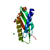
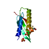
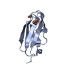
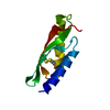
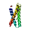
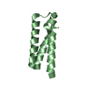
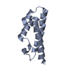
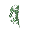
Crystal structure of a fragment of a putative type I restriction enzyme R protein from Bacteroides fragilis
Components
Putative type I restriction enzyme R protein
Keywords
HYDROLASE / STRUCTURAL GENOMICS / UNKNOWN FUNCTION / PSI-2 / Protein Structure Initiative / New York SGX Research Center for Structural Genomics / NYSGXRC
Function / homology
Function and homology information
type I site-specific deoxyribonuclease / type I site-specific deoxyribonuclease activity / DNA restriction-modification system / DNA binding / ATP binding Similarity search - Function
Methane Monooxygenase Hydroxylase; Chain G, domain 1 - #910 / Restriction endonuclease, type I, HsdR, N-terminal / Type I restriction enzyme R protein, C-terminal / SWI2/SNF2 ATPase / : / : / Type I restriction enzyme R protein N terminus (HSDR_N) / Type I restriction and modification enzyme - subunit R C terminal / SWI2/SNF2 ATPase / Type I restriction enzyme subunit R domain 3 ...Methane Monooxygenase Hydroxylase; Chain G, domain 1 - #910 / Restriction endonuclease, type I, HsdR, N-terminal / Type I restriction enzyme R protein, C-terminal / SWI2/SNF2 ATPase / : / : / Type I restriction enzyme R protein N terminus (HSDR_N) / Type I restriction and modification enzyme - subunit R C terminal / SWI2/SNF2 ATPase / Type I restriction enzyme subunit R domain 3 / Restriction endonuclease, type I, HsdR / Methane Monooxygenase Hydroxylase; Chain G, domain 1 / Superfamilies 1 and 2 helicase ATP-binding type-1 domain profile. / DEAD-like helicases superfamily / Helicase superfamily 1/2, ATP-binding domain / Up-down Bundle / P-loop containing nucleoside triphosphate hydrolase / Mainly Alpha Similarity search - Domain/homology
Mass: 18.015 Da / Num. of mol.: 109 / Source method: isolated from a natural source / Formula: H2O
Sequence details
AUTHOR STATE THAT IT IS POSSIBLE THAT WHAT CRYSTALLIZED WAS A PROTEOLYTIC FRAGMENT. THE MS DID NOT ...AUTHOR STATE THAT IT IS POSSIBLE THAT WHAT CRYSTALLIZED WAS A PROTEOLYTIC FRAGMENT. THE MS DID NOT INDICATE A FRAGMENT OF THE APPROXIMATE SIZE OF THE OBSERVED PORTION SO IT IS POSSIBLE FRAGMENTATION OCCURRED DURING THE CRYSTALLIZATION EXPERIMENT. THEREFORE, THE WHOLE SEQUENCE IS LEFT IN THE RECORD.
-
Experimental details
-
Experiment
Experiment
Method: X-RAY DIFFRACTION / Number of used crystals: 1
In the structure databanks used in Yorodumi, some data are registered as the other names, "COVID-19 virus" and "2019-nCoV". Here are the details of the virus and the list of structure data.
Jan 31, 2019. EMDB accession codes are about to change! (news from PDBe EMDB page)
EMDB accession codes are about to change! (news from PDBe EMDB page)
The allocation of 4 digits for EMDB accession codes will soon come to an end. Whilst these codes will remain in use, new EMDB accession codes will include an additional digit and will expand incrementally as the available range of codes is exhausted. The current 4-digit format prefixed with “EMD-” (i.e. EMD-XXXX) will advance to a 5-digit format (i.e. EMD-XXXXX), and so on. It is currently estimated that the 4-digit codes will be depleted around Spring 2019, at which point the 5-digit format will come into force.
The EM Navigator/Yorodumi systems omit the EMD- prefix.
Related info.:Q: What is EMD? / ID/Accession-code notation in Yorodumi/EM Navigator
Yorodumi is a browser for structure data from EMDB, PDB, SASBDB, etc.
This page is also the successor to EM Navigator detail page, and also detail information page/front-end page for Omokage search.
The word "yorodu" (or yorozu) is an old Japanese word meaning "ten thousand". "mi" (miru) is to see.
Related info.:EMDB / PDB / SASBDB / Comparison of 3 databanks / Yorodumi Search / Aug 31, 2016. New EM Navigator & Yorodumi / Yorodumi Papers / Jmol/JSmol / Function and homology information / Changes in new EM Navigator and Yorodumi
 Movie
Movie Controller

 Yorodumi
 Open data
 Basic information
 Components
 Keywords
 Function and homology information
 Bacteroides fragilis NCTC 9343 (bacteria)
 X-RAY DIFFRACTION /
  Authors
 Citation
 Structure visualization
 Downloads & links
 Other downloads


 PDBj
PDBj Assembly
 Mass: 18.015 Da / Num. of mol.: 109 / Source method: isolated from a natural source / Formula: H2O
 Sample preparation
 / Beamline: 31-ID / Wavelength: 0.97958 Å
 Processing