| Entry | Database: PDB / ID: 3et6
|
|---|
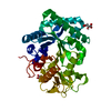
| Title | The crystal structure of the catalytic domain of a eukaryotic guanylate cyclase |
|---|
 Components Components | (Soluble guanylyl cyclase beta) x 2 |
|---|
 Keywords Keywords | LYASE / guanylate cyclase / guanylyl cyclase / dimethylarsenic / Membrane / Transmembrane |
|---|
| Function / homology |  Function and homology information Function and homology information
guanylate cyclase complex, soluble / guanylate cyclase / guanylate cyclase activity / response to oxygen levels / : / heme binding / GTP bindingSimilarity search - Function Haem NO binding associated / Haem NO binding associated domain superfamily / Heme NO binding associated / Heme NO-binding / H-NOX domain superfamily / Haem-NO-binding / Nucleotide cyclase, GGDEF domain / NO signalling/Golgi transport ligand-binding domain superfamily / Adenylyl cyclase class-4/guanylyl cyclase, conserved site / Guanylate cyclase signature. ...Haem NO binding associated / Haem NO binding associated domain superfamily / Heme NO binding associated / Heme NO-binding / H-NOX domain superfamily / Haem-NO-binding / Nucleotide cyclase, GGDEF domain / NO signalling/Golgi transport ligand-binding domain superfamily / Adenylyl cyclase class-4/guanylyl cyclase, conserved site / Guanylate cyclase signature. / Adenylyl- / guanylyl cyclase, catalytic domain / Adenylate and Guanylate cyclase catalytic domain / Adenylyl cyclase class-3/4/guanylyl cyclase / Guanylate cyclase domain profile. / Nucleotide cyclase / Alpha-Beta Plaits / 2-Layer Sandwich / Alpha BetaSimilarity search - Domain/homology |
|---|
| Biological species |   Chlamydomonas reinhardtii (plant) Chlamydomonas reinhardtii (plant) |
|---|
| Method |  X-RAY DIFFRACTION / SYNCHROTRON / MOLECULAR REPLACEMENT / molecular replacement / Resolution: 2.55 Å X-RAY DIFFRACTION / SYNCHROTRON / MOLECULAR REPLACEMENT / molecular replacement / Resolution: 2.55 Å |
|---|
 Authors Authors | Winger, J.A. / Derbyshire, E.R. / Lamers, M.H. / Marletta, M.A. / Kuriyan, J. |
|---|
 Citation Citation | Journal: Bmc Struct.Biol. / Year: 2008
Title: The crystal structure of the catalytic domain of a eukaryotic guanylate cyclase.
Authors: Winger, J.A. / Derbyshire, E.R. / Lamers, M.H. / Marletta, M.A. / Kuriyan, J. |
|---|
| History | | Deposition | Oct 7, 2008 | Deposition site: RCSB / Processing site: RCSB |
|---|
| Revision 1.0 | Oct 14, 2008 | Provider: repository / Type: Initial release |
|---|
| Revision 1.1 | Jul 13, 2011 | Group: Version format compliance |
|---|
| Revision 1.2 | Sep 6, 2023 | Group: Data collection / Database references ...Data collection / Database references / Derived calculations / Refinement description
Category: chem_comp_atom / chem_comp_bond ...chem_comp_atom / chem_comp_bond / database_2 / pdbx_initial_refinement_model / struct_conn / struct_ref_seq_dif / struct_site
Item: _database_2.pdbx_DOI / _database_2.pdbx_database_accession ..._database_2.pdbx_DOI / _database_2.pdbx_database_accession / _struct_conn.pdbx_leaving_atom_flag / _struct_ref_seq_dif.details / _struct_site.pdbx_auth_asym_id / _struct_site.pdbx_auth_comp_id / _struct_site.pdbx_auth_seq_id |
|---|
| Revision 1.3 | Nov 20, 2024 | Group: Structure summary / Category: pdbx_entry_details / pdbx_modification_feature |
|---|
|
|---|
 Movie
Movie Controller

 Yorodumi
 Open data
 Basic information
 Structure visualization
 Downloads & links
 Other downloads


 PDBj
PDBj


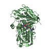
 Assembly


 Mass: 94.971 Da / Num. of mol.: 8 / Source method: obtained synthetically / Formula: PO4
 Mass: 18.015 Da / Num. of mol.: 99 / Source method: isolated from a natural source / Formula: H2O
 Sample preparation
 / Beamline: 8.2.2 / Wavelength: 1 Å
 Processing