 Movie
Movie Controller


+ Open data

- Basic information
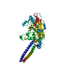
| Entry | Database: PDB / ID: 3esw | |||||||||
|---|---|---|---|---|---|---|---|---|---|---|
| Title | Complex of yeast PNGase with GlcNAc2-IAc. | |||||||||
 Components Components |
| |||||||||
 Keywords Keywords | HYDROLASE / Glycoproteins Peptide:N-glycanase Chitobiose / Metal-binding / Nucleus / DNA damage / DNA repair / Phosphoprotein / Ubl conjugation pathway | |||||||||
| Function / homology |  Function and homology information Function and homology informationubiquitin-dependent glycoprotein ERAD pathway / PNGase complex / peptide-N4-(N-acetyl-beta-glucosaminyl)asparagine amidase / peptide-N4-(N-acetyl-beta-glucosaminyl)asparagine amidase activity / nucleotide-excision repair factor 2 complex / nucleotide-excision repair, DNA damage recognition / K48-linked polyubiquitin modification-dependent protein binding / proteasome binding / protein quality control for misfolded or incompletely synthesized proteins / polyubiquitin modification-dependent protein binding ...ubiquitin-dependent glycoprotein ERAD pathway / PNGase complex / peptide-N4-(N-acetyl-beta-glucosaminyl)asparagine amidase / peptide-N4-(N-acetyl-beta-glucosaminyl)asparagine amidase activity / nucleotide-excision repair factor 2 complex / nucleotide-excision repair, DNA damage recognition / K48-linked polyubiquitin modification-dependent protein binding / proteasome binding / protein quality control for misfolded or incompletely synthesized proteins / polyubiquitin modification-dependent protein binding / ERAD pathway / ubiquitin binding / damaged DNA binding / proteasome-mediated ubiquitin-dependent protein catabolic process / protein-macromolecule adaptor activity / negative regulation of transcription by RNA polymerase II / mitochondrion / nucleoplasm / metal ion binding / nucleus / cytoplasm / cytosol Similarity search - Function | |||||||||
| Biological species |  | |||||||||
| Method |  X-RAY DIFFRACTION / SYNCHROTRON / Resolution: 3.4 Å X-RAY DIFFRACTION / SYNCHROTRON / Resolution: 3.4 Å | |||||||||
 Authors Authors | Zhao, G. / Zhou, X. / Lennarz, W.J. / Schindelin, H. | |||||||||
 Citation Citation | Journal: Glycobiology / Year: 2009 Title: Structural and mutational studies on the importance of oligosaccharide binding for the activity of yeast PNGase. Authors: Zhao, G. / Li, G. / Zhou, X. / Matsuo, I. / Ito, Y. / Suzuki, T. / Lennarz, W.J. / Schindelin, H. | |||||||||
| History |
|
- Structure visualization
| Structure viewer | Molecule: MolmilJmol/JSmol |
|---|
- Downloads & links
-Download
| PDBx/mmCIF format | 3esw.cif.gz | 96.2 KB | Display | PDBx/mmCIF format |
|---|---|---|---|---|
| PDB format | pdb3esw.ent.gz | 71.8 KB | Display | PDB format |
| PDBx/mmJSON format | 3esw.json.gz | Tree view | PDBx/mmJSON format | |
| Others |  Other downloads Other downloads |
-Validation report
| Arichive directory | https://data.pdbj.org/pub/pdb/validation_reports/es/3eswftp://data.pdbj.org/pub/pdb/validation_reports/es/3esw | HTTPS FTP |
|---|
-Related structure data
| Related structure data |  1x3zS S: Starting model for refinement |
|---|---|
| Similar structure data |


-Links
 PDBj
PDBj


- Assembly
| Deposited unit | 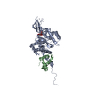
| ||||||||
|---|---|---|---|---|---|---|---|---|---|
| 1 |
| ||||||||
| Unit cell |
|
-Components
| #1: Protein | Mass: 41698.070 Da / Num. of mol.: 1 / Fragment: peptide:N-glycanase Source method: isolated from a genetically manipulated source Source: (gene. exp.) Gene: PNG1, YPL096W / Production host:  References: UniProt: Q02890, peptide-N4-(N-acetyl-beta-glucosaminyl)asparagine amidase |
|---|---|
| #2: Protein | Mass: 6049.967 Da / Num. of mol.: 1 / Fragment: XPCB Domain Source method: isolated from a genetically manipulated source Source: (gene. exp.) Gene: RAD23, SYGP-ORF29, YEL037C / Production host: |
| #3: Polysaccharide | 2-acetamido-2-deoxy-beta-D-glucopyranose-(1-4)-2-acetamido-2-deoxy-beta-D-glucopyranose Source method: isolated from a genetically manipulated source |
| #4: Chemical | ChemComp-ZN /   Mass: 65.409 Da / Num. of mol.: 1 / Source method: obtained synthetically / Formula: Zn Mass: 65.409 Da / Num. of mol.: 1 / Source method: obtained synthetically / Formula: Zn |
| #5: Water | ChemComp-HOH /  Mass: 18.015 Da / Num. of mol.: 7 / Source method: isolated from a natural source / Formula: H2O Mass: 18.015 Da / Num. of mol.: 7 / Source method: isolated from a natural source / Formula: H2O |
-Experimental details
-Experiment
| Experiment | Method: X-RAY DIFFRACTION / Number of used crystals: 1 |
|---|
- Sample preparation
| Crystal | Density Matthews: 6.68 Å3/Da / Density % sol: 81.59 % |
|---|---|
| Crystal grow | Temperature: 291 K / Method: evaporation / pH: 8.5 Details: 0.1 M Tris-HCl, pH 8.5 and 2.0 M sodium chloride, EVAPORATION, temperature 291K |
-Data collection
| Diffraction | Mean temperature: 100 K |
|---|---|
| Diffraction source | Source: SYNCHROTRON / Site: NSLS  / Beamline: X26C / Wavelength: 1 Å / Beamline: X26C / Wavelength: 1 Å |
| Detector | Type: ADSC QUANTUM 4 / Detector: CCD / Date: Aug 19, 2006 |
| Radiation | Protocol: SINGLE WAVELENGTH / Monochromatic (M) / Laue (L): M / Scattering type: x-ray |
| Radiation wavelength | Wavelength: 1 Å / Relative weight: 1 |
| Reflection | Resolution: 3.4→37.969 Å / Num. obs: 17986 / % possible obs: 99.9 % / Observed criterion σ(I): 2 / Redundancy: 8 % / Rmerge(I) obs: 0.126 / Rsym value: 0.126 / Net I/σ(I): 5.496 |
| Reflection shell | Resolution: 3.4→3.58 Å / Redundancy: 8.1 % / Rmerge(I) obs: 0.457 / Mean I/σ(I) obs: 4.4 / Num. measured all: 20859 / Num. unique all: 2581 / Rsym value: 0.457 / % possible all: 100 |
- Processing
| Software |
| ||||||||||||||||||||||||||||||||||||||||||||||||||||||||||||||||||||||||||||||||||||||||||||||||||||
|---|---|---|---|---|---|---|---|---|---|---|---|---|---|---|---|---|---|---|---|---|---|---|---|---|---|---|---|---|---|---|---|---|---|---|---|---|---|---|---|---|---|---|---|---|---|---|---|---|---|---|---|---|---|---|---|---|---|---|---|---|---|---|---|---|---|---|---|---|---|---|---|---|---|---|---|---|---|---|---|---|---|---|---|---|---|---|---|---|---|---|---|---|---|---|---|---|---|---|---|---|---|
| Refinement | Starting model: PDB ENTRY 1X3Z Resolution: 3.4→37.96 Å / Cor.coef. Fo:Fc: 0.949 / Cor.coef. Fo:Fc free: 0.929 / Occupancy max: 1 / Occupancy min: 1 / SU B: 31.04 / SU ML: 0.235 / TLS residual ADP flag: LIKELY RESIDUAL / Cross valid method: THROUGHOUT / σ(F): 0 / ESU R: 0.42 / ESU R Free: 0.304 / Stereochemistry target values: MAXIMUM LIKELIHOOD Details: HYDROGENS HAVE BEEN ADDED IN THE RIDING POSITIONS U VALUES : RESIDUAL ONLY
| ||||||||||||||||||||||||||||||||||||||||||||||||||||||||||||||||||||||||||||||||||||||||||||||||||||
| Solvent computation | Ion probe radii: 0.8 Å / Shrinkage radii: 0.8 Å / VDW probe radii: 1.4 Å / Solvent model: MASK | ||||||||||||||||||||||||||||||||||||||||||||||||||||||||||||||||||||||||||||||||||||||||||||||||||||
| Displacement parameters | Biso max: 106.09 Å2 / Biso mean: 63.61 Å2 / Biso min: 7.26 Å2
| ||||||||||||||||||||||||||||||||||||||||||||||||||||||||||||||||||||||||||||||||||||||||||||||||||||
| Refinement step | Cycle: LAST / Resolution: 3.4→37.96 Å
| ||||||||||||||||||||||||||||||||||||||||||||||||||||||||||||||||||||||||||||||||||||||||||||||||||||
| Refine LS restraints |
| ||||||||||||||||||||||||||||||||||||||||||||||||||||||||||||||||||||||||||||||||||||||||||||||||||||
| LS refinement shell | Resolution: 3.4→3.58 Å / Total num. of bins used: 20
| ||||||||||||||||||||||||||||||||||||||||||||||||||||||||||||||||||||||||||||||||||||||||||||||||||||
| Refinement TLS params. | Method: refined / Refine-ID: X-RAY DIFFRACTION
| ||||||||||||||||||||||||||||||||||||||||||||||||||||||||||||||||||||||||||||||||||||||||||||||||||||
| Refinement TLS group |
|
