 Movie
Movie Controller

[English] 日本語
 Yorodumi
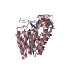
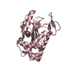
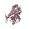
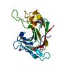
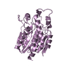
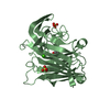
- PDB-3erj: Crystal structure of the peptidyl-tRNA hydrolase AF2095 from Arch... -
+ Open data

- Basic information
| Entry | Database: PDB / ID: 3erj | ||||||
|---|---|---|---|---|---|---|---|
| Title | Crystal structure of the peptidyl-tRNA hydrolase AF2095 from Archaeglobus fulgidis. Northeast Structural Genomics Consortium target GR4 | ||||||
 Components Components | Peptidyl-tRNA hydrolase | ||||||
 Keywords Keywords | HYDROLASE / alpha-beta / Structural Genomics / PSI-2 / Protein Structure Initiative / Northeast Structural Genomics Consortium / NESG / Cytoplasm | ||||||
| Function / homology |  Function and homology information Function and homology informationpeptidyl-tRNA hydrolase / peptidyl-tRNA hydrolase activity / translation / cytosol Similarity search - Function | ||||||
| Biological species |   Archaeoglobus fulgidus (archaea) Archaeoglobus fulgidus (archaea) | ||||||
| Method |  X-RAY DIFFRACTION / SYNCHROTRON / MOLECULAR REPLACEMENT / Resolution: 1.8 Å X-RAY DIFFRACTION / SYNCHROTRON / MOLECULAR REPLACEMENT / Resolution: 1.8 Å | ||||||
 Authors Authors | Forouhar, F. / Su, M. / Seetharaman, J. / Conover, K. / Janjua, H. / Xiao, R. / Cunningham, K. / Ma, L.-C. / Cooper, B. / Baran, M.C. ...Forouhar, F. / Su, M. / Seetharaman, J. / Conover, K. / Janjua, H. / Xiao, R. / Cunningham, K. / Ma, L.-C. / Cooper, B. / Baran, M.C. / Liu, J. / Acton, T.B. / Montelione, G.T. / Tong, L. / Hunt, J.F. / Northeast Structural Genomics Consortium (NESG) | ||||||
 Citation Citation | Journal: To be Published Title: Crystal structure of the peptidyl-tRNA hydrolase AF2095 from Archaeglobus fulgidis. Northeast Structural Genomics Consortium target GR4 Authors: Forouhar, F. / Su, M. / Seetharaman, J. / Conover, K. / Janjua, H. / Xiao, R. / Cunningham, K. / Ma, L.-C. / Cooper, B. / Baran, M.C. / Liu, J. / Acton, T.B. / Montelione, G.T. / Tong, L. / Hunt, J.F. | ||||||
| History |
|
- Structure visualization
| Structure viewer | Molecule: MolmilJmol/JSmol |
|---|
- Downloads & links
-Download
| PDBx/mmCIF format | 3erj.cif.gz | 58.3 KB | Display | PDBx/mmCIF format |
|---|---|---|---|---|
| PDB format | pdb3erj.ent.gz | 42.5 KB | Display | PDB format |
| PDBx/mmJSON format | 3erj.json.gz | Tree view | PDBx/mmJSON format | |
| Others |  Other downloads Other downloads |
-Validation report
| Summary document | 3erj_validation.pdf.gz | 437.9 KB | Display | wwPDB validaton report |
|---|---|---|---|---|
| Full document | 3erj_full_validation.pdf.gz | 441.2 KB | Display | |
| Data in XML | 3erj_validation.xml.gz | 12.3 KB | Display | |
| Data in CIF | 3erj_validation.cif.gz | 16.8 KB | Display | |
| Arichive directory | https://data.pdbj.org/pub/pdb/validation_reports/er/3erjftp://data.pdbj.org/pub/pdb/validation_reports/er/3erj | HTTPS FTP |
-Related structure data
| Related structure data |  1rlkS S: Starting model for refinement |
|---|---|
| Similar structure data | |
| Other databases |


-Links
 PDBj
PDBj- Assembly
| Deposited unit | 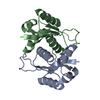
| ||||||||
|---|---|---|---|---|---|---|---|---|---|
| 1 |
| ||||||||
| Unit cell |
|
-Components
| #1: Protein | Mass: 13578.943 Da / Num. of mol.: 2 Source method: isolated from a genetically manipulated source Source: (gene. exp.) Archaeoglobus fulgidus (archaea) / Strain: DSM 4304 / Gene: AF2095, AF_2095, pth / Plasmid: pET21 / Production host:  #2: Water | ChemComp-HOH / |  Mass: 18.015 Da / Num. of mol.: 152 / Source method: isolated from a natural source / Formula: H2O Mass: 18.015 Da / Num. of mol.: 152 / Source method: isolated from a natural source / Formula: H2O |
|---|
-Experimental details
-Experiment
| Experiment | Method: X-RAY DIFFRACTION / Number of used crystals: 1 |
|---|
- Sample preparation
| Crystal | Density Matthews: 2 Å3/Da / Density % sol: 38.38 % |
|---|---|
| Crystal grow | Temperature: 291 K / Method: vapor diffusion, sitting drop / pH: 8 Details: Protein solution: 10 mM Tris (pH 7.5), 100 mM sodium chloride, and 5 mM DTT. Reservoir solution: 100 mM Tris (pH 8), 20% PEG3350, 200 mM NaCl, and 0.5% w/v polyvinylpyrrolidone K15 , VAPOR ...Details: Protein solution: 10 mM Tris (pH 7.5), 100 mM sodium chloride, and 5 mM DTT. Reservoir solution: 100 mM Tris (pH 8), 20% PEG3350, 200 mM NaCl, and 0.5% w/v polyvinylpyrrolidone K15 , VAPOR DIFFUSION, SITTING DROP, temperature 291K |
-Data collection
| Diffraction | Mean temperature: 100 K |
|---|---|
| Diffraction source | Source: SYNCHROTRON / Site: NSLS  / Beamline: X4C / Wavelength: 0.97877 Å / Beamline: X4C / Wavelength: 0.97877 Å |
| Detector | Type: MAR CCD 165 mm / Detector: CCD / Date: Sep 30, 2008 / Details: mirrors |
| Radiation | Monochromator: Si 111 CHANNEL / Protocol: SINGLE WAVELENGTH / Monochromatic (M) / Laue (L): M / Scattering type: x-ray |
| Radiation wavelength | Wavelength: 0.97877 Å / Relative weight: 1 |
| Reflection | Resolution: 1.8→30 Å / Num. all: 38938 / Num. obs: 38783 / % possible obs: 99.6 % / Observed criterion σ(F): 0 / Observed criterion σ(I): 0 / Redundancy: 7.2 % / Biso Wilson estimate: 15 Å2 / Rmerge(I) obs: 0.055 / Rsym value: 0.043 / Net I/σ(I): 34.46 |
| Reflection shell | Resolution: 1.8→1.86 Å / Redundancy: 6.1 % / Rmerge(I) obs: 0.282 / Mean I/σ(I) obs: 4.45 / Num. unique all: 3798 / Rsym value: 0.233 / % possible all: 97.5 |
- Processing
| Software | Name: CNS / Version: 1.2 / Classification: refinement | |||||||||||||||||||||||||
|---|---|---|---|---|---|---|---|---|---|---|---|---|---|---|---|---|---|---|---|---|---|---|---|---|---|---|
| Refinement | Method to determine structure: MOLECULAR REPLACEMENT Starting model: 1RLK Resolution: 1.8→19.88 Å / Rfactor Rfree error: 0.005 / Data cutoff high absF: 114421.04 / Data cutoff low absF: 0 / Isotropic thermal model: RESTRAINED / Cross valid method: THROUGHOUT / σ(F): 2 / σ(I): 2 / Stereochemistry target values: Engh & Huber
| |||||||||||||||||||||||||
| Solvent computation | Solvent model: FLAT MODEL / Bsol: 61.7469 Å2 / ksol: 0.4 e/Å3 | |||||||||||||||||||||||||
| Displacement parameters | Biso mean: 24.3 Å2
| |||||||||||||||||||||||||
| Refine analyze |
| |||||||||||||||||||||||||
| Refinement step | Cycle: LAST / Resolution: 1.8→19.88 Å
| |||||||||||||||||||||||||
| Refine LS restraints |
| |||||||||||||||||||||||||
| LS refinement shell | Resolution: 1.8→1.86 Å / Rfactor Rfree error: 0.021 / Total num. of bins used: 10
|
