[English] 日本語
Yorodumi
- PDB-3eft: Crystal structure of the complex between Carbonic Anhydrase II an... -

+
Open data


ID or keywords:

Loading...

-
Basic information

Entry
Database: PDB / ID: 3eft
TitleCrystal structure of the complex between Carbonic Anhydrase II and a spin-labeled sulfonamide incorporating TEMPO moiety
ComponentsCarbonic anhydrase 2
KeywordsLYASE / carbonic anhydrase / inhibitors / Radicals / OXO-ACID
Function / homology
Function and homology information


positive regulation of cellular pH reduction / positive regulation of dipeptide transmembrane transport / regulation of monoatomic anion transport / secretion / cyanamide hydratase / cyanamide hydratase activity / arylesterase activity / regulation of chloride transport / Reversible hydration of carbon dioxide / angiotensin-activated signaling pathway ...positive regulation of cellular pH reduction / positive regulation of dipeptide transmembrane transport / regulation of monoatomic anion transport / secretion / cyanamide hydratase / cyanamide hydratase activity / arylesterase activity / regulation of chloride transport / Reversible hydration of carbon dioxide / angiotensin-activated signaling pathway / positive regulation of synaptic transmission, GABAergic / regulation of intracellular pH / morphogenesis of an epithelium / carbonic anhydrase / carbonate dehydratase activity / carbon dioxide transport / Erythrocytes take up oxygen and release carbon dioxide / Erythrocytes take up carbon dioxide and release oxygen / neuron cellular homeostasis / one-carbon metabolic process / apical part of cell / myelin sheath / extracellular exosome / zinc ion binding / plasma membrane / cytosol / cytoplasm
Similarity search - Function
Carbonic Anhydrase II / Alpha carbonic anhydrase / Carbonic anhydrase, alpha-class, conserved site / Alpha-carbonic anhydrases signature. / Carbonic anhydrase, alpha-class / Alpha carbonic anhydrase domain / Alpha carbonic anhydrase domain superfamily / Eukaryotic-type carbonic anhydrase / Alpha-carbonic anhydrases profile. / Eukaryotic-type carbonic anhydrase ...Carbonic Anhydrase II / Alpha carbonic anhydrase / Carbonic anhydrase, alpha-class, conserved site / Alpha-carbonic anhydrases signature. / Carbonic anhydrase, alpha-class / Alpha carbonic anhydrase domain / Alpha carbonic anhydrase domain superfamily / Eukaryotic-type carbonic anhydrase / Alpha-carbonic anhydrases profile. / Eukaryotic-type carbonic anhydrase / Roll / Alpha Beta
Similarity search - Domain/homology
Chem-3BS / : / Carbonic anhydrase 2
Similarity search - Component
MethodX-RAY DIFFRACTION / MOLECULAR REPLACEMENT / Resolution: 1.85 Å
AuthorsTemperini, C. / Cecchi, A. / Scozzafava, A. / Supuran, C.T.
CitationJournal: J.Phys.Chem.B / Year: 2009
Title: Dissecting the Inhibition Mechanism of Cytosolic versus Transmembrane Carbonic Anhydrases by ESR
Authors: Ciani, L. / Cecchi, A. / Temperini, C. / Supuran, C.T. / Ristori, S.
History
DepositionSep 10, 2008Deposition site: RCSB / Processing site: PDBJ
Revision 1.0Sep 8, 2009Provider: repository / Type: Initial release
Revision 1.1Jul 13, 2011Group: Version format compliance
Revision 1.2Oct 25, 2017Group: Refinement description / Category: software / Item: _software.name
Revision 1.3Nov 1, 2023Group: Data collection / Database references ...Data collection / Database references / Derived calculations / Refinement description
Category: chem_comp_atom / chem_comp_bond ...chem_comp_atom / chem_comp_bond / database_2 / pdbx_initial_refinement_model / struct_site
Item: _database_2.pdbx_DOI / _database_2.pdbx_database_accession ..._database_2.pdbx_DOI / _database_2.pdbx_database_accession / _struct_site.pdbx_auth_asym_id / _struct_site.pdbx_auth_comp_id / _struct_site.pdbx_auth_seq_id

-
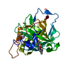
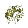
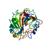
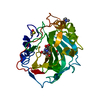
Structure visualization

Structure viewerMolecule:
MolmilJmol/JSmol

Downloads & links

-
Assembly

Deposited unit
A: Carbonic anhydrase 2
hetero molecules


Theoretical massNumber of molelcules
Total (without water)29,9764
Polymers29,2891
Non-polymers6873
Water3,909217
1


  • Idetical with deposited unit
  • defined by author&software
TypeNameSymmetry operationNumber
identity operation1_555x,y,z1
MethodPISA
Unit cell
Length a, b, c (Å)42.130, 41.500, 72.310
Angle α, β, γ (deg.)90.00, 104.30, 90.00
Int Tables number4
Space group name H-MP1211

-
Components

#1: Protein Carbonic anhydrase 2 / / Carbonic anhydrase II / CA-II / Carbonate dehydratase II / Carbonic anhydrase C / CAC


Mass: 29289.062 Da / Num. of mol.: 1 / Source method: obtained synthetically
Details: Chemically synthesized. This sequence occurs naturally in humans(homo sapiens).
References: UniProt: P00918, carbonic anhydrase
#2: Chemical ChemComp-ZN / ZINC ION


Mass: 65.409 Da / Num. of mol.: 1 / Source method: obtained synthetically / Formula: Zn
#3: Chemical ChemComp-HG / MERCURY (II) ION / Mercury (element)


Mass: 200.590 Da / Num. of mol.: 1 / Source method: obtained synthetically / Formula: Hg
#4: Chemical ChemComp-3BS / 3-chloro-4-{[(1-hydroxy-2,2,6,6-tetramethylpiperidin-4-yl)carbamothioyl]amino}benzenesulfonamide / (4-{[(2-chloro-4-sulfamoylphenyl)carbamothioyl]amino}-2,2,6,6-tetramethylpiperidin-1-yl)oxidanyl


Mass: 420.978 Da / Num. of mol.: 1 / Source method: obtained synthetically / Formula: C16H25ClN4O3S2
#5: Water ChemComp-HOH / water / Water


Mass: 18.015 Da / Num. of mol.: 217 / Source method: isolated from a natural source / Formula: H2O
Sequence detailsTHE NUMBER 126 IS SIMPLY SKIPPED IN THE NUMBERING. THE GAP IS IN THE SEQUENCE NUMBERING OF MODEL ...THE NUMBER 126 IS SIMPLY SKIPPED IN THE NUMBERING. THE GAP IS IN THE SEQUENCE NUMBERING OF MODEL 1CA2, THAT IS THE STARTING MODEL FOR ALL CA2 STRUCTURES.

-
Experimental details

-
Experiment

ExperimentMethod: X-RAY DIFFRACTION / Number of used crystals: 1

-
Sample preparation

CrystalDensity Matthews: 2.09 Å3/Da / Density % sol: 41.19 %
Crystal growTemperature: 277 K / Method: vapor diffusion, hanging drop
Details: 50mM Tris.HCl pH 7.7-7.8, 2mM sodium 4-(hydroxymercury)benzoate, VAPOR DIFFUSION, HANGING DROP, temperature 277K
PH range: 7.7-7.8

-
Data collection

DiffractionMean temperature: 100 K
Diffraction sourceSource: SEALED TUBE / Type: OXFORD DIFFRACTION ENHANCE ULTRA / Wavelength: 1.5418 Å
DetectorType: OXFORD SAPPHIRE CCD / Detector: CCD / Date: Jun 1, 2008
RadiationMonochromator: capillary / Protocol: SINGLE WAVELENGTH / Monochromatic (M) / Laue (L): M / Scattering type: x-ray
Radiation wavelengthWavelength: 1.5418 Å / Relative weight: 1
ReflectionResolution: 1.84→20 Å / Num. all: 20861 / Num. obs: 20988 / % possible obs: 89.4 % / Observed criterion σ(F): 2 / Observed criterion σ(I): 0 / Redundancy: 2.9 % / Rsym value: 0.09 / Net I/σ(I): 13.1

-
Processing

Software
NameVersionClassification
CrysalisProOxford Diffraction2006data collection
AMoREphasing
REFMAC5.2.0019refinement
CrysalisProOxford Diffraction2006data reduction
CrysalisProOxford Diffraction2006data scaling
RefinementMethod to determine structure: MOLECULAR REPLACEMENT
Starting model: PDB ENTRY 1CA2
Resolution: 1.85→11.81 Å / Cor.coef. Fo:Fc: 0.938 / Cor.coef. Fo:Fc free: 0.914 / SU B: 3.553 / SU ML: 0.108 / Cross valid method: THROUGHOUT / ESU R: 0.171 / ESU R Free: 0.155 / Stereochemistry target values: MAXIMUM LIKELIHOOD / Details: HYDROGENS HAVE BEEN ADDED IN THE RIDING POSITIONS
RfactorNum. reflection% reflectionSelection details
Rfree0.2403 1040 5 %RANDOM
Rwork0.19231 ---
obs0.19469 19758 99.5 %-
Solvent computationIon probe radii: 0.8 Å / Shrinkage radii: 0.8 Å / VDW probe radii: 1.2 Å / Solvent model: MASK
Displacement parametersBiso mean: 13.824 Å2
Baniso -1Baniso -2Baniso -3
1-0 Å20 Å20.06 Å2
2--0.16 Å20 Å2
3----0.12 Å2
Refinement stepCycle: LAST / Resolution: 1.85→11.81 Å
ProteinNucleic acidLigandSolventTotal
Num. atoms2078 0 28 217 2323
Refine LS restraints
Refine-IDTypeDev idealDev ideal targetNumber
X-RAY DIFFRACTIONr_bond_refined_d0.0090.0222177
X-RAY DIFFRACTIONr_angle_refined_deg1.2891.9662978
X-RAY DIFFRACTIONr_dihedral_angle_1_deg5.9425274
X-RAY DIFFRACTIONr_dihedral_angle_2_deg35.61124.74799
X-RAY DIFFRACTIONr_dihedral_angle_3_deg16.36615360
X-RAY DIFFRACTIONr_dihedral_angle_4_deg18.228157
X-RAY DIFFRACTIONr_chiral_restr0.2090.2314
X-RAY DIFFRACTIONr_gen_planes_refined0.0040.021682
X-RAY DIFFRACTIONr_nbd_refined0.1980.21037
X-RAY DIFFRACTIONr_nbtor_refined0.3050.21429
X-RAY DIFFRACTIONr_xyhbond_nbd_refined0.1240.2207
X-RAY DIFFRACTIONr_metal_ion_refined0.0630.23
X-RAY DIFFRACTIONr_symmetry_vdw_refined0.1730.252
X-RAY DIFFRACTIONr_symmetry_hbond_refined0.10.218
X-RAY DIFFRACTIONr_mcbond_it0.7161.51347
X-RAY DIFFRACTIONr_mcangle_it0.92322119
X-RAY DIFFRACTIONr_scbond_it1.5863969
X-RAY DIFFRACTIONr_scangle_it2.3414.5849
LS refinement shellResolution: 1.85→1.898 Å / Total num. of bins used: 20
RfactorNum. reflection% reflection
Rfree0.282 71 -
Rwork0.238 1461 -
obs--100 %

+
About Yorodumi

-
News

-
Feb 9, 2022. New format data for meta-information of EMDB entries

New format data for meta-information of EMDB entries

  • Version 3 of the EMDB header file is now the official format.
  • The previous official version 1.9 will be removed from the archive.

Related info.:EMDB header

External links:wwPDB to switch to version 3 of the EMDB data model

-
Aug 12, 2020. Covid-19 info

Covid-19 info

URL: https://pdbj.org/emnavi/covid19.php

New page: Covid-19 featured information page in EM Navigator.

Related info.:Covid-19 info / Mar 5, 2020. Novel coronavirus structure data

+
Mar 5, 2020. Novel coronavirus structure data

Novel coronavirus structure data

Related info.:Yorodumi Speices / Aug 12, 2020. Covid-19 info

External links:COVID-19 featured content - PDBj / Molecule of the Month (242):Coronavirus Proteases

+
Jan 31, 2019. EMDB accession codes are about to change! (news from PDBe EMDB page)

EMDB accession codes are about to change! (news from PDBe EMDB page)

  • The allocation of 4 digits for EMDB accession codes will soon come to an end. Whilst these codes will remain in use, new EMDB accession codes will include an additional digit and will expand incrementally as the available range of codes is exhausted. The current 4-digit format prefixed with “EMD-” (i.e. EMD-XXXX) will advance to a 5-digit format (i.e. EMD-XXXXX), and so on. It is currently estimated that the 4-digit codes will be depleted around Spring 2019, at which point the 5-digit format will come into force.
  • The EM Navigator/Yorodumi systems omit the EMD- prefix.

Related info.:Q: What is EMD? / ID/Accession-code notation in Yorodumi/EM Navigator

External links:EMDB Accession Codes are Changing Soon! / Contact to PDBj

+
Jul 12, 2017. Major update of PDB

Major update of PDB

  • wwPDB released updated PDB data conforming to the new PDBx/mmCIF dictionary.
  • This is a major update changing the version number from 4 to 5, and with Remediation, in which all the entries are updated.
  • In this update, many items about electron microscopy experimental information are reorganized (e.g. em_software).
  • Now, EM Navigator and Yorodumi are based on the updated data.

External links:wwPDB Remediation / Enriched Model Files Conforming to OneDep Data Standards Now Available in the PDB FTP Archive

-
Yorodumi

Thousand views of thousand structures

  • Yorodumi is a browser for structure data from EMDB, PDB, SASBDB, etc.
  • This page is also the successor to EM Navigator detail page, and also detail information page/front-end page for Omokage search.
  • The word "yorodu" (or yorozu) is an old Japanese word meaning "ten thousand". "mi" (miru) is to see.

Related info.:EMDB / PDB / SASBDB / Comparison of 3 databanks / Yorodumi Search / Aug 31, 2016. New EM Navigator & Yorodumi / Yorodumi Papers / Jmol/JSmol / Function and homology information / Changes in new EM Navigator and Yorodumi

Read more