- PDB-3ef0: The Structure of Fcp1, an essential RNA polymerase II CTD phosphatase -
+
Open data
ID or keywords:
Loading...
-
Basic information
Entry
Database: PDB / ID: 3ef0
Title
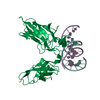
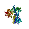
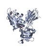
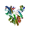
The Structure of Fcp1, an essential RNA polymerase II CTD phosphatase
Components
RNA polymerase II subunit A C-terminal domain phosphatase
Keywords
HYDROLASE / phosphatase / CTD / FCPH / BRCT / AlF4 / transition state analog / Cobalt / Magnesium / Manganese / Metal-binding / Nucleus / Protein phosphatase
Function / homology
Function and homology information
Formation of the Early Elongation Complex / RNA Polymerase II Pre-transcription Events / TP53 Regulates Transcription of DNA Repair Genes / RNA polymerase II CTD heptapeptide repeat phosphatase activity / signaling / protein-serine/threonine phosphatase / metal ion binding / nucleus Similarity search - Function
Mass: 18.015 Da / Num. of mol.: 175 / Source method: isolated from a natural source / Formula: H2O
Sequence details
UNP Q9P376 RESIDUES 330-393 ARE DELETED AND REPLACED BY RESIDUES 'SG'
-
Experimental details
-
Experiment
Experiment
Method: X-RAY DIFFRACTION / Number of used crystals: 1
-
Sample preparation
Crystal
Density Matthews: 2.35 Å3/Da / Density % sol: 47.56 %
Crystal grow
Temperature: 291 K / Method: vapor diffusion, hanging drop / pH: 7 Details: Fcp1-AlF4--Mg was obtained by incubating 250 M Fcp1(149-580)- 330-393 with 500 M AlCl3, 5 mM NaF and 5 mM MgCl2 on ice for 2 h prior to crystallization by hanging drop vapor diffusion ...Details: Fcp1-AlF4--Mg was obtained by incubating 250 M Fcp1(149-580)- 330-393 with 500 M AlCl3, 5 mM NaF and 5 mM MgCl2 on ice for 2 h prior to crystallization by hanging drop vapor diffusion against 20% PEG-3350, 190 mM ammonium formate (pH 7.0), VAPOR DIFFUSION, HANGING DROP, temperature 291K
In the structure databanks used in Yorodumi, some data are registered as the other names, "COVID-19 virus" and "2019-nCoV". Here are the details of the virus and the list of structure data.
Jan 31, 2019. EMDB accession codes are about to change! (news from PDBe EMDB page)
EMDB accession codes are about to change! (news from PDBe EMDB page)
The allocation of 4 digits for EMDB accession codes will soon come to an end. Whilst these codes will remain in use, new EMDB accession codes will include an additional digit and will expand incrementally as the available range of codes is exhausted. The current 4-digit format prefixed with “EMD-” (i.e. EMD-XXXX) will advance to a 5-digit format (i.e. EMD-XXXXX), and so on. It is currently estimated that the 4-digit codes will be depleted around Spring 2019, at which point the 5-digit format will come into force.
The EM Navigator/Yorodumi systems omit the EMD- prefix.
Related info.:Q: What is EMD? / ID/Accession-code notation in Yorodumi/EM Navigator
Yorodumi is a browser for structure data from EMDB, PDB, SASBDB, etc.
This page is also the successor to EM Navigator detail page, and also detail information page/front-end page for Omokage search.
The word "yorodu" (or yorozu) is an old Japanese word meaning "ten thousand". "mi" (miru) is to see.
Related info.:EMDB / PDB / SASBDB / Comparison of 3 databanks / Yorodumi Search / Aug 31, 2016. New EM Navigator & Yorodumi / Yorodumi Papers / Jmol/JSmol / Function and homology information / Changes in new EM Navigator and Yorodumi
 Movie
Movie Controller

 Yorodumi
 Open data
 Basic information
 Components
 Keywords
 Function and homology information
 X-RAY DIFFRACTION /
  Authors
 Citation
 Structure visualization
 Downloads & links
 Other downloads


 PDBj
PDBj


 Assembly


 Mass: 102.975 Da / Num. of mol.: 1 / Source method: obtained synthetically / Formula: AlF4
 Mass: 24.305 Da / Num. of mol.: 1 / Source method: obtained synthetically / Formula: Mg
 Mass: 18.015 Da / Num. of mol.: 175 / Source method: isolated from a natural source / Formula: H2O
 Sample preparation
 / Beamline: 24-ID-C / Wavelength: 0.979 Å
 Processing