 Movie
Movie Controller

[English] 日本語
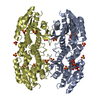
 Yorodumi
- PDB-3edm: Crystal structure of a short chain dehydrogenase from Agrobacteri... -
+ Open data

- Basic information
| Entry | Database: PDB / ID: 3edm | ||||||
|---|---|---|---|---|---|---|---|
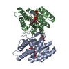
| Title | Crystal structure of a short chain dehydrogenase from Agrobacterium tumefaciens | ||||||
 Components Components | Short chain dehydrogenase | ||||||
 Keywords Keywords | OXIDOREDUCTASE / STRUCTURAL GENOMICS / DEHYDROGENASE / PSI-2 / Protein Structure Initiative / New York SGX Research Center for Structural Genomics / NYSGXRC | ||||||
| Function / homology |  Function and homology information Function and homology information | ||||||
| Biological species |  Agrobacterium tumefaciens str. (bacteria) Agrobacterium tumefaciens str. (bacteria) | ||||||
| Method |  X-RAY DIFFRACTION / SYNCHROTRON / SAD / Resolution: 2.3 Å X-RAY DIFFRACTION / SYNCHROTRON / SAD / Resolution: 2.3 Å | ||||||
 Authors Authors | Bonanno, J.B. / Rutter, M. / Bain, K.T. / Chang, S. / Romero, R. / Wasserman, S. / Sauder, J.M. / Burley, S.K. / Almo, S.C. / New York SGX Research Center for Structural Genomics (NYSGXRC) | ||||||
 Citation Citation | Journal: To be Published Title: Crystal structure of a short chain dehydrogenase from Agrobacterium tumefaciens Authors: Bonanno, J.B. / Rutter, M. / Bain, K.T. / Chang, S. / Romero, R. / Wasserman, S. / Sauder, J.M. / Burley, S.K. / Almo, S.C. | ||||||
| History |
|
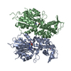
- Structure visualization
| Structure viewer | Molecule: MolmilJmol/JSmol |
|---|
- Downloads & links
-Download
| PDBx/mmCIF format | 3edm.cif.gz | 166.2 KB | Display | PDBx/mmCIF format |
|---|---|---|---|---|
| PDB format | pdb3edm.ent.gz | 132.3 KB | Display | PDB format |
| PDBx/mmJSON format | 3edm.json.gz | Tree view | PDBx/mmJSON format | |
| Others |  Other downloads Other downloads |
-Validation report
| Arichive directory | https://data.pdbj.org/pub/pdb/validation_reports/ed/3edmftp://data.pdbj.org/pub/pdb/validation_reports/ed/3edm | HTTPS FTP |
|---|
-Related structure data
| Similar structure data | |
|---|---|
| Other databases |


-Links
 PDBj
PDBj

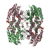
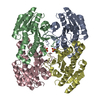
- Assembly
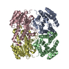
| Deposited unit | 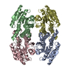
| ||||||||
|---|---|---|---|---|---|---|---|---|---|
| 1 |
| ||||||||
| Unit cell |
|
-Components
| #1: Protein | Mass: 26911.521 Da / Num. of mol.: 4 Source method: isolated from a genetically manipulated source Source: (gene. exp.) Agrobacterium tumefaciens str. (bacteria)Strain: C58 / Gene: AGR_L_3556, Atu3026 / Plasmid: modified pET26 / Production host: #2: Water | ChemComp-HOH / |  Mass: 18.015 Da / Num. of mol.: 189 / Source method: isolated from a natural source / Formula: H2O Mass: 18.015 Da / Num. of mol.: 189 / Source method: isolated from a natural source / Formula: H2O |
|---|
-Experimental details
-Experiment
| Experiment | Method: X-RAY DIFFRACTION / Number of used crystals: 1 |
|---|
- Sample preparation
| Crystal | Density Matthews: 2.37 Å3/Da / Density % sol: 48.14 % |
|---|---|
| Crystal grow | Temperature: 294 K / Method: vapor diffusion / pH: 7 Details: 22% PEG 3350, 200mM sodium formate, pH 7.0, Vapor diffusion, temperature 294K |
-Data collection
| Diffraction | Mean temperature: 100 K |
|---|---|
| Diffraction source | Source: SYNCHROTRON / Site: ALS  / Beamline: 8.3.1 / Wavelength: 0.97958 Å / Beamline: 8.3.1 / Wavelength: 0.97958 Å |
| Detector | Type: ADSC QUANTUM 315 / Detector: CCD / Date: Aug 28, 2008 |
| Radiation | Monochromator: Si 111 / Protocol: SINGLE WAVELENGTH / Monochromatic (M) / Laue (L): M / Scattering type: x-ray |
| Radiation wavelength | Wavelength: 0.97958 Å / Relative weight: 1 |
| Reflection | Resolution: 2.3→43.783 Å / Num. all: 46266 / Num. obs: 46266 / % possible obs: 100 % / Observed criterion σ(F): 0 / Observed criterion σ(I): 0 / Redundancy: 8 % / Biso Wilson estimate: 33.8 Å2 / Rmerge(I) obs: 0.099 / Rsym value: 0.099 / Net I/σ(I): 15.3 |
| Reflection shell | Resolution: 2.3→2.42 Å / Redundancy: 8.2 % / Rmerge(I) obs: 0.472 / Mean I/σ(I) obs: 4.5 / Num. measured all: 54360 / Num. unique all: 6669 / Rsym value: 0.472 / % possible all: 100 |
- Processing
| Software |
| |||||||||||||||||||||||||||||||||||||||||||||||||||||||||||||||||
|---|---|---|---|---|---|---|---|---|---|---|---|---|---|---|---|---|---|---|---|---|---|---|---|---|---|---|---|---|---|---|---|---|---|---|---|---|---|---|---|---|---|---|---|---|---|---|---|---|---|---|---|---|---|---|---|---|---|---|---|---|---|---|---|---|---|---|
| Refinement | Method to determine structure: SAD / Resolution: 2.3→20 Å / Cor.coef. Fo:Fc: 0.959 / Cor.coef. Fo:Fc free: 0.928 / WRfactor Rfree: 0.241 / WRfactor Rwork: 0.181 / Occupancy max: 1 / Occupancy min: 0.5 / FOM work R set: 0.776 / SU B: 6.627 / SU ML: 0.163 / SU R Cruickshank DPI: 0.273 / SU Rfree: 0.233 / Cross valid method: THROUGHOUT / σ(F): 0 / σ(I): 0 / ESU R: 0.273 / ESU R Free: 0.233 / Stereochemistry target values: MAXIMUM LIKELIHOOD / Details: HYDROGENS HAVE BEEN ADDED IN THE RIDING POSITIONS
| |||||||||||||||||||||||||||||||||||||||||||||||||||||||||||||||||
| Solvent computation | Ion probe radii: 0.8 Å / Shrinkage radii: 0.8 Å / VDW probe radii: 1.2 Å / Solvent model: BABINET MODEL WITH MASK | |||||||||||||||||||||||||||||||||||||||||||||||||||||||||||||||||
| Displacement parameters | Biso max: 70.01 Å2 / Biso mean: 35.109 Å2 / Biso min: 18.3 Å2
| |||||||||||||||||||||||||||||||||||||||||||||||||||||||||||||||||
| Refinement step | Cycle: LAST / Resolution: 2.3→20 Å
| |||||||||||||||||||||||||||||||||||||||||||||||||||||||||||||||||
| Refine LS restraints |
| |||||||||||||||||||||||||||||||||||||||||||||||||||||||||||||||||
| LS refinement shell | Resolution: 2.3→2.359 Å / Total num. of bins used: 20
|
