[English] 日本語
Yorodumi
- PDB-3eck: Structure of E323L Homoprotocatechuate 2,3-dioxygenase from Brevi... -

+
Open data


ID or keywords:

Loading...

-
Basic information

Entry
Database: PDB / ID: 3eck
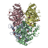
Title
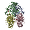
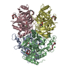
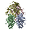
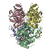
Structure of E323L Homoprotocatechuate 2,3-dioxygenase from Brevibacterium fuscum in complex with putative O-O bond cleavage intermediate formed via in crystallo reaction with 4-sulfonyl catechol at low oxygen concentrations
ComponentsPROTEIN (Homoprotocatechuate 2,3-dioxygenase)
KeywordsOXIDOREDUCTASE / oxygenase / extradiol / FeII / crystal packing / Dioxygenase
Function / homology
Function and homology information


dioxygenase activity / metal ion binding
Similarity search - Function
homoprotocatechuate 2,3-dioxygenase fold / homoprotocatechuate 2,3-dioxygenase domains / 3,4-dihydroxyphenylacetate 2,3-dioxygenase, Mn/Fe-type / 3,4-dihydroxyphenylacetate 2,3-dioxygenase, C-terminal domain superfamily / 2,3-Dihydroxybiphenyl 1,2-Dioxygenase, domain 1 / 2,3-Dihydroxybiphenyl 1,2-Dioxygenase; domain 1 / Glyoxalase/fosfomycin resistance/dioxygenase domain / Glyoxalase/Bleomycin resistance protein/Dioxygenase superfamily / Vicinal oxygen chelate (VOC) domain / Vicinal oxygen chelate (VOC) domain profile. ...homoprotocatechuate 2,3-dioxygenase fold / homoprotocatechuate 2,3-dioxygenase domains / 3,4-dihydroxyphenylacetate 2,3-dioxygenase, Mn/Fe-type / 3,4-dihydroxyphenylacetate 2,3-dioxygenase, C-terminal domain superfamily / 2,3-Dihydroxybiphenyl 1,2-Dioxygenase, domain 1 / 2,3-Dihydroxybiphenyl 1,2-Dioxygenase; domain 1 / Glyoxalase/fosfomycin resistance/dioxygenase domain / Glyoxalase/Bleomycin resistance protein/Dioxygenase superfamily / Vicinal oxygen chelate (VOC) domain / Vicinal oxygen chelate (VOC) domain profile. / Glyoxalase/Bleomycin resistance protein/Dihydroxybiphenyl dioxygenase / Few Secondary Structures / Irregular / Roll / Alpha Beta
Similarity search - Domain/homology
: / Chem-XXG / Homoprotocatechuate 2,3-dioxygenase
Similarity search - Component
Biological speciesBrevibacterium fuscum (bacteria)
MethodX-RAY DIFFRACTION / SYNCHROTRON / MOLECULAR REPLACEMENT / Resolution: 1.6 Å
AuthorsKovaleva, E.G. / Lipscomb, J.D.
CitationJournal: Biochemistry / Year: 2008
Title: Intermediate in the O-O Bond Cleavage Reaction of an Extradiol Dioxygenase.
Authors: Kovaleva, E.G. / Lipscomb, J.D.
History
DepositionAug 31, 2008Deposition site: RCSB / Processing site: RCSB
Revision 1.0Oct 7, 2008Provider: repository / Type: Initial release
Revision 1.1Jul 13, 2011Group: Non-polymer description / Version format compliance
Revision 1.2Oct 20, 2021Group: Database references / Derived calculations
Category: database_2 / pdbx_struct_conn_angle ...database_2 / pdbx_struct_conn_angle / struct_conn / struct_ref_seq_dif / struct_site
Item: _database_2.pdbx_DOI / _database_2.pdbx_database_accession ..._database_2.pdbx_DOI / _database_2.pdbx_database_accession / _pdbx_struct_conn_angle.ptnr1_auth_seq_id / _pdbx_struct_conn_angle.ptnr1_label_alt_id / _pdbx_struct_conn_angle.ptnr3_auth_seq_id / _pdbx_struct_conn_angle.ptnr3_label_alt_id / _pdbx_struct_conn_angle.value / _struct_conn.pdbx_dist_value / _struct_conn.pdbx_ptnr2_label_alt_id / _struct_conn.ptnr1_auth_asym_id / _struct_conn.ptnr1_auth_comp_id / _struct_conn.ptnr1_auth_seq_id / _struct_conn.ptnr1_label_asym_id / _struct_conn.ptnr1_label_atom_id / _struct_conn.ptnr1_label_comp_id / _struct_conn.ptnr1_label_seq_id / _struct_conn.ptnr2_auth_asym_id / _struct_conn.ptnr2_auth_comp_id / _struct_conn.ptnr2_auth_seq_id / _struct_conn.ptnr2_label_asym_id / _struct_conn.ptnr2_label_atom_id / _struct_conn.ptnr2_label_comp_id / _struct_ref_seq_dif.details / _struct_site.pdbx_auth_asym_id / _struct_site.pdbx_auth_comp_id / _struct_site.pdbx_auth_seq_id
Revision 1.3Aug 30, 2023Group: Data collection / Refinement description
Category: chem_comp_atom / chem_comp_bond / pdbx_initial_refinement_model

-
Structure visualization

Structure viewerMolecule:
MolmilJmol/JSmol

Downloads & links

-
Assembly

Deposited unit
A: PROTEIN (Homoprotocatechuate 2,3-dioxygenase)
B: PROTEIN (Homoprotocatechuate 2,3-dioxygenase)
C: PROTEIN (Homoprotocatechuate 2,3-dioxygenase)
D: PROTEIN (Homoprotocatechuate 2,3-dioxygenase)
hetero molecules


Theoretical massNumber of molelcules
Total (without water)168,32821
Polymers166,9574
Non-polymers1,37017
Water25,0951393
1


  • Idetical with deposited unit
  • defined by author&software
TypeNameSymmetry operationNumber
identity operation1_555x,y,z1
Buried area16760 Å2
ΔGint-105 kcal/mol
Surface area43620 Å2
MethodPISA
Unit cell
Length a, b, c (Å)110.735, 163.176, 101.373
Angle α, β, γ (deg.)90.00, 90.00, 90.00
Int Tables number18
Space group name H-MP21212

-
Components

-
Protein , 1 types, 4 molecules ABCD

#1: Protein
PROTEIN (Homoprotocatechuate 2,3-dioxygenase)


Mass: 41739.363 Da / Num. of mol.: 4 / Mutation: E323L
Source method: isolated from a genetically manipulated source
Source: (gene. exp.) Brevibacterium fuscum (bacteria) / Strain: ATCC 15993 / Gene: hpcd / Plasmid: pYZW204 / Production host: Escherichia coli (E. coli) / Strain (production host): BL21(DE3)
References: UniProt: Q45135, 3,4-dihydroxyphenylacetate 2,3-dioxygenase

-
Non-polymers , 6 types, 1410 molecules

#2: Chemical
ChemComp-FE2 / FE (II) ION


Mass: 55.845 Da / Num. of mol.: 4 / Source method: obtained synthetically / Formula: Fe
#3: Chemical
ChemComp-CL / CHLORIDE ION / Chloride


Mass: 35.453 Da / Num. of mol.: 4 / Source method: obtained synthetically / Formula: Cl
#4: Chemical
ChemComp-GOL / GLYCEROL / GLYCERIN / PROPANE-1,2,3-TRIOL / Glycerol


Mass: 92.094 Da / Num. of mol.: 6 / Source method: obtained synthetically / Formula: C3H8O3
#5: Chemical ChemComp-CA / CALCIUM ION


Mass: 40.078 Da / Num. of mol.: 1 / Source method: obtained synthetically / Formula: Ca
#6: Chemical ChemComp-XXG / 3,3-dihydroxy-4-oxocyclohexa-1,5-diene-1-sulfonic acid / 1,1-dihydroxy-2-keto-5-sulfonyl-cyclohexa-3,5-diene


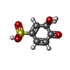
Mass: 206.173 Da / Num. of mol.: 2 / Source method: obtained synthetically / Formula: C6H6O6S
#7: Water ChemComp-HOH / water / Water


Mass: 18.015 Da / Num. of mol.: 1393 / Source method: isolated from a natural source / Formula: H2O

-
Experimental details

-
Experiment

ExperimentMethod: X-RAY DIFFRACTION / Number of used crystals: 1

-
Sample preparation

CrystalDensity Matthews: 2.74 Å3/Da / Density % sol: 55.16 %
Crystal growTemperature: 292 K / Method: vapor diffusion, hanging drop / pH: 7
Details: 18% PEG8000, 0.1M Ca acetate, 0.1M Na cacodylate, pH 7.0, VAPOR DIFFUSION, HANGING DROP, temperature 292K

-
Data collection

DiffractionMean temperature: 100 K
Diffraction sourceSource: SYNCHROTRON / Site: APS / Beamline: 19-BM / Wavelength: 0.97903 Å
DetectorType: SBC-3 / Detector: CCD / Date: Jun 17, 2007
RadiationProtocol: SINGLE WAVELENGTH / Monochromatic (M) / Laue (L): M / Scattering type: x-ray
Radiation wavelengthWavelength: 0.97903 Å / Relative weight: 1
ReflectionResolution: 1.6→55 Å / Num. all: 240583 / Num. obs: 222399 / % possible obs: 92.4 % / Observed criterion σ(F): 0 / Observed criterion σ(I): -3 / Redundancy: 4.1 % / Rmerge(I) obs: 0.052 / Rsym value: 0.052 / Net I/σ(I): 19.22
Reflection shellResolution: 1.6→1.66 Å / Redundancy: 3.3 % / Rmerge(I) obs: 0.342 / Mean I/σ(I) obs: 3.067 / Num. unique all: 17442 / % possible all: 73.2

-
Processing

Software
NameVersionClassification
HKL-3000data collection
MOLREPphasing
REFMAC5.2.0019refinement
HKL-3000data reduction
HKL-3000data scaling
RefinementMethod to determine structure: MOLECULAR REPLACEMENT
Starting model: PDB ID 2IG9
Resolution: 1.6→24.2 Å / Cor.coef. Fo:Fc: 0.968 / Cor.coef. Fo:Fc free: 0.96 / SU B: 1.553 / SU ML: 0.054 / Cross valid method: THROUGHOUT / ESU R: 0.085 / ESU R Free: 0.083 / Stereochemistry target values: MAXIMUM LIKELIHOOD / Details: HYDROGENS HAVE BEEN ADDED IN THE RIDING POSITIONS
RfactorNum. reflection% reflectionSelection details
Rfree0.19767 11135 5 %RANDOM
Rwork0.17401 ---
obs0.17521 211226 92.41 %-
Solvent computationIon probe radii: 0.8 Å / Shrinkage radii: 0.8 Å / VDW probe radii: 1.4 Å / Solvent model: MASK
Displacement parametersBiso mean: 19.045 Å2
Baniso -1Baniso -2Baniso -3
1-0 Å20 Å20 Å2
2--0 Å20 Å2
3---0 Å2
Refinement stepCycle: LAST / Resolution: 1.6→24.2 Å
ProteinNucleic acidLigandSolventTotal
Num. atoms11596 0 71 1393 13060
Refine LS restraints
Refine-IDTypeDev idealDev ideal targetNumber
X-RAY DIFFRACTIONr_bond_refined_d0.0110.02112264
X-RAY DIFFRACTIONr_angle_refined_deg1.2971.94716720
X-RAY DIFFRACTIONr_dihedral_angle_1_deg6.70451518
X-RAY DIFFRACTIONr_dihedral_angle_2_deg32.27723.463667
X-RAY DIFFRACTIONr_dihedral_angle_3_deg12.249151961
X-RAY DIFFRACTIONr_dihedral_angle_4_deg16.86615108
X-RAY DIFFRACTIONr_chiral_restr0.0950.21753
X-RAY DIFFRACTIONr_gen_planes_refined0.0060.029764
X-RAY DIFFRACTIONr_nbd_refined0.20.25983
X-RAY DIFFRACTIONr_nbtor_refined0.3040.28397
X-RAY DIFFRACTIONr_xyhbond_nbd_refined0.110.21360
X-RAY DIFFRACTIONr_metal_ion_refined0.1180.22
X-RAY DIFFRACTIONr_symmetry_vdw_refined0.1770.249
X-RAY DIFFRACTIONr_symmetry_hbond_refined0.1970.220
X-RAY DIFFRACTIONr_symmetry_metal_ion_refined0.0970.22
X-RAY DIFFRACTIONr_mcbond_it0.71.57501
X-RAY DIFFRACTIONr_mcangle_it1.078211870
X-RAY DIFFRACTIONr_scbond_it1.76935371
X-RAY DIFFRACTIONr_scangle_it2.7154.54825
LS refinement shellResolution: 1.601→1.642 Å / Total num. of bins used: 20
RfactorNum. reflection% reflection
Rfree0.352 598 -
Rwork0.292 11609 -
obs--69.64 %

+
About Yorodumi

-
News

-
Feb 9, 2022. New format data for meta-information of EMDB entries

New format data for meta-information of EMDB entries

  • Version 3 of the EMDB header file is now the official format.
  • The previous official version 1.9 will be removed from the archive.

Related info.:EMDB header

External links:wwPDB to switch to version 3 of the EMDB data model

-
Aug 12, 2020. Covid-19 info

Covid-19 info

URL: https://pdbj.org/emnavi/covid19.php

New page: Covid-19 featured information page in EM Navigator.

Related info.:Covid-19 info / Mar 5, 2020. Novel coronavirus structure data

+
Mar 5, 2020. Novel coronavirus structure data

Novel coronavirus structure data

Related info.:Yorodumi Speices / Aug 12, 2020. Covid-19 info

External links:COVID-19 featured content - PDBj / Molecule of the Month (242):Coronavirus Proteases

+
Jan 31, 2019. EMDB accession codes are about to change! (news from PDBe EMDB page)

EMDB accession codes are about to change! (news from PDBe EMDB page)

  • The allocation of 4 digits for EMDB accession codes will soon come to an end. Whilst these codes will remain in use, new EMDB accession codes will include an additional digit and will expand incrementally as the available range of codes is exhausted. The current 4-digit format prefixed with “EMD-” (i.e. EMD-XXXX) will advance to a 5-digit format (i.e. EMD-XXXXX), and so on. It is currently estimated that the 4-digit codes will be depleted around Spring 2019, at which point the 5-digit format will come into force.
  • The EM Navigator/Yorodumi systems omit the EMD- prefix.

Related info.:Q: What is EMD? / ID/Accession-code notation in Yorodumi/EM Navigator

External links:EMDB Accession Codes are Changing Soon! / Contact to PDBj

+
Jul 12, 2017. Major update of PDB

Major update of PDB

  • wwPDB released updated PDB data conforming to the new PDBx/mmCIF dictionary.
  • This is a major update changing the version number from 4 to 5, and with Remediation, in which all the entries are updated.
  • In this update, many items about electron microscopy experimental information are reorganized (e.g. em_software).
  • Now, EM Navigator and Yorodumi are based on the updated data.

External links:wwPDB Remediation / Enriched Model Files Conforming to OneDep Data Standards Now Available in the PDB FTP Archive

-
Yorodumi

Thousand views of thousand structures

  • Yorodumi is a browser for structure data from EMDB, PDB, SASBDB, etc.
  • This page is also the successor to EM Navigator detail page, and also detail information page/front-end page for Omokage search.
  • The word "yorodu" (or yorozu) is an old Japanese word meaning "ten thousand". "mi" (miru) is to see.

Related info.:EMDB / PDB / SASBDB / Comparison of 3 databanks / Yorodumi Search / Aug 31, 2016. New EM Navigator & Yorodumi / Yorodumi Papers / Jmol/JSmol / Function and homology information / Changes in new EM Navigator and Yorodumi

Read more