 Movie
Movie Controller

[English] 日本語
 Yorodumi
- PDB-3e8v: Crystal structure of a possible transglutaminase-family protein p... -
+ Open data

- Basic information
| Entry | Database: PDB / ID: 3e8v | ||||||
|---|---|---|---|---|---|---|---|
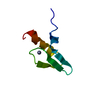
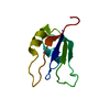
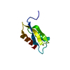
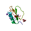
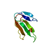
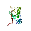
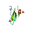
| Title | Crystal structure of a possible transglutaminase-family protein proteolytic fragment from Bacteroides fragilis | ||||||
 Components Components | Possible transglutaminase-family protein | ||||||
 Keywords Keywords | structural genomics / unknown function / PSI-2 / Protein Structure Initiative / New York SGX Research Center for Structural Genomics / NYSGXRC | ||||||
| Function / homology |  Function and homology information Function and homology informationCarboxypeptidase-like, regulatory domain / Transglutaminase-like superfamily / Transglutaminase/protease-like homologues / Transglutaminase-like / Papain-like cysteine peptidase superfamily / Prokaryotic membrane lipoprotein lipid attachment site profile. / Immunoglobulin-like / Sandwich / Mainly Beta Similarity search - Domain/homology | ||||||
| Biological species |  Bacteroides fragilis NCTC 9343 (bacteria) Bacteroides fragilis NCTC 9343 (bacteria) | ||||||
| Method |  X-RAY DIFFRACTION / SYNCHROTRON / SAD / Resolution: 2.4 Å X-RAY DIFFRACTION / SYNCHROTRON / SAD / Resolution: 2.4 Å | ||||||
 Authors Authors | Bonanno, J.B. / Rutter, M. / Bain, K.T. / Hu, S. / Romero, R. / Smith, D. / Wasserman, S. / Sauder, J.M. / Burley, S.K. / Almo, S.C. / New York SGX Research Center for Structural Genomics (NYSGXRC) | ||||||
 Citation Citation | Journal: To be Published Title: Crystal structure of a possible transglutaminase-family protein proteolytic fragment from Bacteroides fragilis Authors: Bonanno, J.B. / Rutter, M. / Bain, K.T. / Hu, S. / Romero, R. / Smith, D. / Wasserman, S. / Sauder, J.M. / Burley, S.K. / Almo, S.C. | ||||||
| History |
|
- Structure visualization
| Structure viewer | Molecule: MolmilJmol/JSmol |
|---|
- Downloads & links
-Download
| PDBx/mmCIF format | 3e8v.cif.gz | 28.7 KB | Display | PDBx/mmCIF format |
|---|---|---|---|---|
| PDB format | pdb3e8v.ent.gz | 18.6 KB | Display | PDB format |
| PDBx/mmJSON format | 3e8v.json.gz | Tree view | PDBx/mmJSON format | |
| Others |  Other downloads Other downloads |
-Validation report
| Arichive directory | https://data.pdbj.org/pub/pdb/validation_reports/e8/3e8vftp://data.pdbj.org/pub/pdb/validation_reports/e8/3e8v | HTTPS FTP |
|---|
-Related structure data
| Similar structure data | |
|---|---|
| Other databases |

-Links
 PDBj
PDBj


- Assembly
| Deposited unit | 
| ||||||||
|---|---|---|---|---|---|---|---|---|---|
| 1 |
| ||||||||
| Unit cell |
| ||||||||
| Components on special symmetry positions |
| ||||||||
| Details | authors state that the biological unit is probable dimer mediated by unknown ion. |

-Components
| #1: Protein | Mass: 8917.956 Da / Num. of mol.: 1 / Fragment: residues 289-370 Source method: isolated from a genetically manipulated source Source: (gene. exp.) Bacteroides fragilis NCTC 9343 (bacteria)Strain: ATCC 25285 / Gene: BF1045 / Plasmid: modified pET26 / Production host: |
|---|---|
| #2: Chemical | ChemComp-UNL / Num. of mol.: 1 / Source method: obtained synthetically |
| #3: Water | ChemComp-HOH /  Mass: 18.015 Da / Num. of mol.: 5 / Source method: isolated from a natural source / Formula: H2O Mass: 18.015 Da / Num. of mol.: 5 / Source method: isolated from a natural source / Formula: H2O |
| Sequence details | AUTHORS STATE THAT THE CRYSTAL IS FORMED FROM A FRAGMENT OF A PROTEIN OF 847 AMINO ACIDS, AND IT IS ...AUTHORS STATE THAT THE CRYSTAL IS FORMED FROM A FRAGMENT OF A PROTEIN OF 847 AMINO ACIDS, AND IT IS UNKNOWN WHAT THE ACTUAL TERMINI ARE FOR THE CRYSTALLIZ |
-Experimental details
-Experiment
| Experiment | Method: X-RAY DIFFRACTION / Number of used crystals: 1 |
|---|
- Sample preparation
| Crystal | Density Matthews: 3.52 Å3/Da / Density % sol: 65.09 % |
|---|---|
| Crystal grow | Temperature: 294 K / Method: vapor diffusion / pH: 6.5 Details: 100mM sodium cacodylate pH 6.5, 1.4M sodium acetate hydrate, Vapor diffusion, temperature 294K |
-Data collection
| Diffraction | Mean temperature: 100 K |
|---|---|
| Diffraction source | Source: SYNCHROTRON / Site: APS  / Beamline: 31-ID / Wavelength: 0.97958 Å / Beamline: 31-ID / Wavelength: 0.97958 Å |
| Detector | Type: MAR CCD 165 mm / Detector: CCD / Date: Jul 18, 2008 |
| Radiation | Monochromator: diamond / Protocol: SINGLE WAVELENGTH / Monochromatic (M) / Laue (L): M / Scattering type: x-ray |
| Radiation wavelength | Wavelength: 0.97958 Å / Relative weight: 1 |
| Reflection | Resolution: 2.4→19.699 Å / Num. all: 5084 / Num. obs: 5079 / % possible obs: 99.9 % / Observed criterion σ(F): 0 / Observed criterion σ(I): 0 / Redundancy: 22 % / Biso Wilson estimate: 51.6 Å2 / Rmerge(I) obs: 0.1 / Rsym value: 0.1 / Net I/σ(I): 30.1 |
| Reflection shell | Resolution: 2.4→2.53 Å / Redundancy: 22.3 % / Rmerge(I) obs: 0.574 / Mean I/σ(I) obs: 5.8 / Num. measured all: 16470 / Num. unique all: 737 / Rsym value: 0.574 / % possible all: 99.8 |
- Processing
| Software |
| |||||||||||||||||||||||||||||||||||||||||||||||||||||||||||||||||||||||||||||||||||||||||||||||
|---|---|---|---|---|---|---|---|---|---|---|---|---|---|---|---|---|---|---|---|---|---|---|---|---|---|---|---|---|---|---|---|---|---|---|---|---|---|---|---|---|---|---|---|---|---|---|---|---|---|---|---|---|---|---|---|---|---|---|---|---|---|---|---|---|---|---|---|---|---|---|---|---|---|---|---|---|---|---|---|---|---|---|---|---|---|---|---|---|---|---|---|---|---|---|---|---|
| Refinement | Method to determine structure: SAD / Resolution: 2.4→15 Å / Cor.coef. Fo:Fc: 0.932 / Cor.coef. Fo:Fc free: 0.924 / WRfactor Rfree: 0.239 / WRfactor Rwork: 0.22 / Occupancy max: 1 / Occupancy min: 0.5 / FOM work R set: 0.82 / SU B: 6.792 / SU ML: 0.155 / SU R Cruickshank DPI: 0.276 / SU Rfree: 0.21 / Cross valid method: THROUGHOUT / σ(F): 0 / σ(I): 0 / ESU R: 0.276 / ESU R Free: 0.21 / Stereochemistry target values: MAXIMUM LIKELIHOOD Details: HYDROGENS HAVE BEEN ADDED IN THE RIDING POSITIONS; UNKNOWN ION MODELED AS BA; PROTEOLYTIC FRAGMENT IDENTIFIED BY SEQUENCE REGISTER
| |||||||||||||||||||||||||||||||||||||||||||||||||||||||||||||||||||||||||||||||||||||||||||||||
| Solvent computation | Ion probe radii: 0.8 Å / Shrinkage radii: 0.8 Å / VDW probe radii: 1.4 Å / Solvent model: MASK | |||||||||||||||||||||||||||||||||||||||||||||||||||||||||||||||||||||||||||||||||||||||||||||||
| Displacement parameters | Biso max: 57.06 Å2 / Biso mean: 36.837 Å2 / Biso min: 18.71 Å2
| |||||||||||||||||||||||||||||||||||||||||||||||||||||||||||||||||||||||||||||||||||||||||||||||
| Refinement step | Cycle: LAST / Resolution: 2.4→15 Å
| |||||||||||||||||||||||||||||||||||||||||||||||||||||||||||||||||||||||||||||||||||||||||||||||
| Refine LS restraints |
| |||||||||||||||||||||||||||||||||||||||||||||||||||||||||||||||||||||||||||||||||||||||||||||||
| LS refinement shell | Resolution: 2.4→2.461 Å / Total num. of bins used: 20
|
