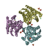
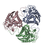
regulation of L-arginine import across plasma membrane / Urea cycle / collagen biosynthetic process / mammary gland involution / positive regulation of neutrophil mediated killing of fungus / arginine metabolic process / negative regulation of T-helper 2 cell cytokine production / arginase / arginase activity / : ...regulation of L-arginine import across plasma membrane / Urea cycle / collagen biosynthetic process / mammary gland involution / positive regulation of neutrophil mediated killing of fungus / arginine metabolic process / negative regulation of T-helper 2 cell cytokine production / arginase / arginase activity / : / response to selenium ion / urea cycle / response to methylmercury / response to nematode / response to manganese ion / response to vitamin A / response to steroid hormone / response to herbicide / Neutrophil degranulation / response to zinc ion / response to vitamin E / defense response to protozoan / response to amine / negative regulation of type II interferon-mediated signaling pathway / negative regulation of activated T cell proliferation / maternal process involved in female pregnancy / cellular response to dexamethasone stimulus / response to cadmium ion / response to amino acid / cellular response to transforming growth factor beta stimulus / response to axon injury / negative regulation of T cell proliferation / positive regulation of endothelial cell proliferation / cellular response to glucagon stimulus / cellular response to interleukin-4 / lung development / liver development / female pregnancy / response to peptide hormone / cellular response to hydrogen peroxide / manganese ion binding / cellular response to lipopolysaccharide / response to lipopolysaccharide / adaptive immune response / mitochondrial outer membrane / response to xenobiotic stimulus / innate immune response / neuronal cell body / extracellular space / identical protein binding / cytosol / cytoplasm 類似検索 - 分子機能
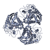
Arginase / Ureohydrolase domain / Ureohydrolase, manganese-binding site / Arginase family signature. / Ureohydrolase / Arginase family / Arginase family profile. / Arginase; Chain A / Ureohydrolase domain superfamily / 3-Layer(aba) Sandwich / Alpha Beta 類似検索 - ドメイン・相同性
手法: 蒸気拡散法, ハンギングドロップ法 詳細: drops containing 3 microL of protein solution [5 mg/mL protein, 50 mM bicine (pH 8.5), 2 mM BEC, 2 mM MnCl2] and 3 microL of precipitant solution [0.1 M CHES (pH 9.5), 20% PEG 3350, 0.2 M ...詳細: drops containing 3 microL of protein solution [5 mg/mL protein, 50 mM bicine (pH 8.5), 2 mM BEC, 2 mM MnCl2] and 3 microL of precipitant solution [0.1 M CHES (pH 9.5), 20% PEG 3350, 0.2 M NaCl] were equilibrated over a 1 mL reservoir of precipitant solution, VAPOR DIFFUSION, HANGING DROP
-
データ収集
回折
平均測定温度: 100 K
放射光源
由来: シンクロトロン / サイト: CHESS / ビームライン: A1 / 波長: 1 Å
検出器
タイプ: ADSC QUANTUM 210 / 検出器: CCD
放射
プロトコル: SINGLE WAVELENGTH / 単色(M)・ラウエ(L): M / 散乱光タイプ: x-ray
放射波長
波長: 1 Å / 相対比: 1
反射
解像度: 2.9→50 Å / Num. all: 18754 / Num. obs: 18754 / % possible obs: 96.3 % / 冗長度: 2.9 % / Biso Wilson estimate: 15.4 Å2 / Rmerge(I) obs: 0.136 / Net I/σ(I): 9.3
反射 シェル
解像度: 2.9→3 Å / 冗長度: 2.7 % / Rmerge(I) obs: 0.33 / Mean I/σ(I) obs: 2.7 / Num. unique all: 1644 / % possible all: 90.5
 ムービー
ムービー コントローラー


 データを開く
 基本情報
基本情報 要素
要素 キーワード
キーワード 機能・相同性情報
 X線回折 /
  データ登録者
 引用
引用 構造の表示
構造の表示 ダウンロードとリンク
 その他のダウンロード
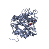


 PDBj
PDBj


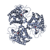
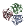
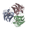
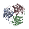
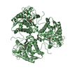
 集合体
集合体


 分子量: 54.938 Da / 分子数: 6 / 由来タイプ: 合成 / 式: Mn
 分子量: 18.015 Da / 分子数: 46 / 由来タイプ: 天然 / 式: H2O
 試料調製
試料調製 / ビームライン: A1 / 波長: 1 Å
 解析
解析