 Movie
Movie Controller


+ Open data

- Basic information
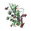
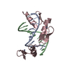
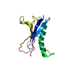
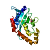
| Entry | Database: PDB / ID: 3e4g | ||||||
|---|---|---|---|---|---|---|---|
| Title | Crystal structure of bovine coupling Factor B, G28E mutant | ||||||
 Components Components | ATP synthase subunit s, mitochondrial | ||||||
 Keywords Keywords | ELECTRON TRANSPORT / leucine-rich repeat / CF0 / Hydrogen ion transport / Inner membrane / Ion transport / Membrane / Mitochondrion / Transit peptide / Transport | ||||||
| Function / homology |  Function and homology information Function and homology informationFormation of ATP by chemiosmotic coupling / Cristae formation / ATP biosynthetic process / proton-transporting ATP synthase complex / proton transmembrane transport / mitochondrial inner membrane / metal ion binding / cytoplasm Similarity search - Function | ||||||
| Biological species |  | ||||||
| Method |  X-RAY DIFFRACTION / SYNCHROTRON / MOLECULAR REPLACEMENT / molecular replacement / Resolution: 0.96 Å X-RAY DIFFRACTION / SYNCHROTRON / MOLECULAR REPLACEMENT / molecular replacement / Resolution: 0.96 Å | ||||||
 Authors Authors | Stroud, R.M. / Lee, J.K. / Belogrudov, G.I. | ||||||
 Citation Citation | Journal: Proc.Natl.Acad.Sci.Usa / Year: 2008 Title: Crystal structure of bovine mitochondrial factor B at 0.96-A resolution. Authors: Lee, J.K. / Belogrudov, G.I. / Stroud, R.M. | ||||||
| History |
|
- Structure visualization
| Structure viewer | Molecule: MolmilJmol/JSmol |
|---|
- Downloads & links
-Download
| PDBx/mmCIF format | 3e4g.cif.gz | 101 KB | Display | PDBx/mmCIF format |
|---|---|---|---|---|
| PDB format | pdb3e4g.ent.gz | 76.1 KB | Display | PDB format |
| PDBx/mmJSON format | 3e4g.json.gz | Tree view | PDBx/mmJSON format | |
| Others |  Other downloads Other downloads |
-Validation report
| Arichive directory | https://data.pdbj.org/pub/pdb/validation_reports/e4/3e4gftp://data.pdbj.org/pub/pdb/validation_reports/e4/3e4g | HTTPS FTP |
|---|
-Related structure data
| Related structure data | 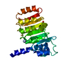 3dzeSC  3e2jC 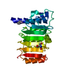 3e3zC S: Starting model for refinement C: citing same article ( |
|---|---|
| Similar structure data |


-Links
 PDBj
PDBj

- Assembly
| Deposited unit | 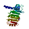
| ||||||||
|---|---|---|---|---|---|---|---|---|---|
| 1 |
| ||||||||
| Unit cell |
|
-Components
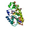
| #1: Protein | Mass: 20514.738 Da / Num. of mol.: 1 / Mutation: G28E Source method: isolated from a genetically manipulated source Source: (gene. exp.)  References: UniProt: P22027, H+-transporting two-sector ATPase |
|---|---|
| #2: Chemical | ChemComp-MG /   Mass: 24.305 Da / Num. of mol.: 1 / Source method: obtained synthetically / Formula: Mg Mass: 24.305 Da / Num. of mol.: 1 / Source method: obtained synthetically / Formula: Mg |
| #3: Chemical | ChemComp-TRS /   Mass: 122.143 Da / Num. of mol.: 1 / Source method: obtained synthetically / Formula: C4H12NO3 / Comment: pH buffer*YM Mass: 122.143 Da / Num. of mol.: 1 / Source method: obtained synthetically / Formula: C4H12NO3 / Comment: pH buffer*YM |
| #4: Water | ChemComp-HOH /  Mass: 18.015 Da / Num. of mol.: 265 / Source method: isolated from a natural source / Formula: H2O Mass: 18.015 Da / Num. of mol.: 265 / Source method: isolated from a natural source / Formula: H2O |
-Experimental details
-Experiment
| Experiment | Method: X-RAY DIFFRACTION / Number of used crystals: 1 |
|---|
- Sample preparation
| Crystal | Density Matthews: 2.28 Å3/Da / Density % sol: 46.04 % |
|---|---|
| Crystal grow | Temperature: 277 K / Method: vapor diffusion / pH: 7.4 Details: 50% PPG 400, 100 mM Tris-HCl pH 7.4, VAPOR DIFFUSION, temperature 277K |
-Data collection
| Diffraction | Mean temperature: 100 K |
|---|---|
| Diffraction source | Source: SYNCHROTRON / Site: ALS  / Beamline: 8.3.1 / Wavelength: 1.0332 Å / Beamline: 8.3.1 / Wavelength: 1.0332 Å |
| Detector | Type: ADSC QUANTUM 210 / Detector: CCD / Date: May 12, 2007 / Details: KOHZU |
| Radiation | Monochromator: Double crystal Si(111) / Protocol: SINGLE WAVELENGTH / Scattering type: x-ray |
| Radiation wavelength | Wavelength: 1.0332 Å / Relative weight: 1 |
| Reflection | Resolution: 0.955→50.7 Å / Num. all: 95943 / Num. obs: 92547 / % possible obs: 99.99 % / Observed criterion σ(I): 2 / Redundancy: 4.2 % / Biso Wilson estimate: 9.763 Å2 / Rmerge(I) obs: 0.078 / Net I/σ(I): 15.7 |
| Reflection shell | Resolution: 0.955→0.985 Å / Redundancy: 1.6 % / Mean I/σ(I) obs: 2 / Num. unique all: 1198 / Rsym value: 0.33 / % possible all: 100 |
-Phasing
| Phasing | Method: molecular replacement | |||||||||
|---|---|---|---|---|---|---|---|---|---|---|
| Phasing MR | Model details: Phaser MODE: MR_AUTO
|
- Processing
| Software |
| ||||||||||||||||||||||||||||||||||||||||||||||||||||||||||||||||||||||||||||||||||||||||||||||||||||
|---|---|---|---|---|---|---|---|---|---|---|---|---|---|---|---|---|---|---|---|---|---|---|---|---|---|---|---|---|---|---|---|---|---|---|---|---|---|---|---|---|---|---|---|---|---|---|---|---|---|---|---|---|---|---|---|---|---|---|---|---|---|---|---|---|---|---|---|---|---|---|---|---|---|---|---|---|---|---|---|---|---|---|---|---|---|---|---|---|---|---|---|---|---|---|---|---|---|---|---|---|---|
| Refinement | Method to determine structure: MOLECULAR REPLACEMENT Starting model: PDB entry 3DZE Resolution: 0.96→30 Å / Cor.coef. Fo:Fc: 0.977 / Cor.coef. Fo:Fc free: 0.971 / WRfactor Rfree: 0.174 / WRfactor Rwork: 0.151 / Occupancy max: 1 / Occupancy min: 0.5 / FOM work R set: 0.924 / SU B: 0.49 / SU ML: 0.012 / SU R Cruickshank DPI: 0.022 / SU Rfree: 0.023 / Cross valid method: THROUGHOUT / σ(F): 0 / ESU R: 0.022 / ESU R Free: 0.022 / Stereochemistry target values: MAXIMUM LIKELIHOOD / Details: HYDROGENS HAVE BEEN ADDED IN THE RIDING POSITIONS
| ||||||||||||||||||||||||||||||||||||||||||||||||||||||||||||||||||||||||||||||||||||||||||||||||||||
| Solvent computation | Ion probe radii: 0.8 Å / Shrinkage radii: 0.8 Å / VDW probe radii: 1.2 Å / Solvent model: MASK | ||||||||||||||||||||||||||||||||||||||||||||||||||||||||||||||||||||||||||||||||||||||||||||||||||||
| Displacement parameters | Biso max: 56.3 Å2 / Biso mean: 14.542 Å2 / Biso min: 5.39 Å2
| ||||||||||||||||||||||||||||||||||||||||||||||||||||||||||||||||||||||||||||||||||||||||||||||||||||
| Refinement step | Cycle: LAST / Resolution: 0.96→30 Å
| ||||||||||||||||||||||||||||||||||||||||||||||||||||||||||||||||||||||||||||||||||||||||||||||||||||
| Refine LS restraints |
| ||||||||||||||||||||||||||||||||||||||||||||||||||||||||||||||||||||||||||||||||||||||||||||||||||||
| LS refinement shell | Resolution: 0.96→0.985 Å / Total num. of bins used: 20
|
