 Movie
Movie Controller

[English] 日本語
 Yorodumi
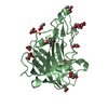
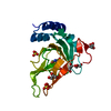
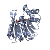
- PDB-3e1g: Snapshots of esterase D from lactobacillus rhamnosus: Insights in... -
+ Open data

- Basic information
| Entry | Database: PDB / ID: 3e1g | ||||||
|---|---|---|---|---|---|---|---|
| Title | Snapshots of esterase D from lactobacillus rhamnosus: Insights into a rotation driven catalytic mechanism | ||||||
 Components Components | Esterase D | ||||||
 Keywords Keywords | HYDROLASE / alpha beta hydrolase / catalytic triad / rotation / esterase | ||||||
| Function / homology |  Function and homology information Function and homology information | ||||||
| Biological species |  Lactobacillus rhamnosus (bacteria) Lactobacillus rhamnosus (bacteria) | ||||||
| Method |  X-RAY DIFFRACTION / MOLECULAR REPLACEMENT / molecular replacement / Resolution: 2.2 Å X-RAY DIFFRACTION / MOLECULAR REPLACEMENT / molecular replacement / Resolution: 2.2 Å | ||||||
 Authors Authors | Bennett, M.D. / Delabre, M.-L. / Holland, R. / Norris, G.E. | ||||||
 Citation Citation | Journal: To be Published Title: Snapshots of esterase D from lactobacillus rhamnosus: Insights into a rotation driven catalytic mechanism Authors: Bennett, M.D. / Delabre, M.-L. / Holland, R. / Norris, G.E. | ||||||
| History |
|
- Structure visualization
| Structure viewer | Molecule: MolmilJmol/JSmol |
|---|
- Downloads & links
-Download
| PDBx/mmCIF format | 3e1g.cif.gz | 59.7 KB | Display | PDBx/mmCIF format |
|---|---|---|---|---|
| PDB format | pdb3e1g.ent.gz | 42.2 KB | Display | PDB format |
| PDBx/mmJSON format | 3e1g.json.gz | Tree view | PDBx/mmJSON format | |
| Others |  Other downloads Other downloads |
-Validation report
| Arichive directory | https://data.pdbj.org/pub/pdb/validation_reports/e1/3e1gftp://data.pdbj.org/pub/pdb/validation_reports/e1/3e1g | HTTPS FTP |
|---|
-Related structure data
| Related structure data |  3dltC  3dyiC  3dyvC  1r1dS C: citing same article ( S: Starting model for refinement |
|---|---|
| Similar structure data |


-Links
 PDBj
PDBj- Assembly
| Deposited unit | 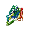
| ||||||||
|---|---|---|---|---|---|---|---|---|---|
| 1 |
| ||||||||
| Unit cell |
|
-Components
| #1: Protein | Mass: 27327.963 Da / Num. of mol.: 1 Source method: isolated from a genetically manipulated source Source: (gene. exp.) Lactobacillus rhamnosus (bacteria) / Strain: HN001 / Gene: EstD / Plasmid: pPROEXHTc / Production host: |
|---|---|
| #2: Chemical | ChemComp-DEP /   Mass: 138.102 Da / Num. of mol.: 1 / Source method: obtained synthetically / Formula: C4H11O3P Mass: 138.102 Da / Num. of mol.: 1 / Source method: obtained synthetically / Formula: C4H11O3P |
| #3: Water | ChemComp-HOH /  Mass: 18.015 Da / Num. of mol.: 87 / Source method: isolated from a natural source / Formula: H2O Mass: 18.015 Da / Num. of mol.: 87 / Source method: isolated from a natural source / Formula: H2O |
-Experimental details
-Experiment
| Experiment | Method: X-RAY DIFFRACTION / Number of used crystals: 1 |
|---|
- Sample preparation
| Crystal | Density Matthews: 2.07 Å3/Da / Density % sol: 40.49 % |
|---|---|
| Crystal grow | Temperature: 298 K / Method: hanging drop / pH: 6 Details: PEG 8000, Na acetate, pH 6.0, hanging drop, temperature 298K |
-Data collection
| Diffraction | Mean temperature: 120 K |
|---|---|
| Diffraction source | Source: ROTATING ANODE / Type: RIGAKU MICROMAX-007 / Wavelength: 1.54 Å |
| Detector | Type: RIGAKU RAXIS IV / Detector: IMAGE PLATE / Date: Oct 30, 2006 / Details: Osmic Blue |
| Radiation | Monochromator: Osmic Blue / Protocol: SINGLE WAVELENGTH / Monochromatic (M) / Laue (L): M / Scattering type: x-ray |
| Radiation wavelength | Wavelength: 1.54 Å / Relative weight: 1 |
| Reflection | Resolution: 1.49→33.52 Å / Num. all: 38533 / Num. obs: 34921 / % possible obs: 90.6 % / Observed criterion σ(F): 0 / Observed criterion σ(I): 0 / Redundancy: 7.6 % / Rmerge(I) obs: 0.157 / Net I/σ(I): 5.1 |
-Phasing
| Phasing | Method: molecular replacement |
|---|
- Processing
| Software |
| |||||||||||||||||||||||||||||||||||||||||||||||||||||||||||||||||||||||||||||||||||||||||||||||||||||||||
|---|---|---|---|---|---|---|---|---|---|---|---|---|---|---|---|---|---|---|---|---|---|---|---|---|---|---|---|---|---|---|---|---|---|---|---|---|---|---|---|---|---|---|---|---|---|---|---|---|---|---|---|---|---|---|---|---|---|---|---|---|---|---|---|---|---|---|---|---|---|---|---|---|---|---|---|---|---|---|---|---|---|---|---|---|---|---|---|---|---|---|---|---|---|---|---|---|---|---|---|---|---|---|---|---|---|---|
| Refinement | Method to determine structure: MOLECULAR REPLACEMENT Starting model: 1R1D Resolution: 2.2→33.52 Å / Cor.coef. Fo:Fc: 0.95 / Cor.coef. Fo:Fc free: 0.914 / WRfactor Rfree: 0.249 / WRfactor Rwork: 0.198 / Occupancy max: 1 / Occupancy min: 1 / FOM work R set: 0.817 / SU B: 6.02 / SU ML: 0.155 / SU R Cruickshank DPI: 0.248 / SU Rfree: 0.224 / Cross valid method: THROUGHOUT / σ(F): 0 / σ(I): 0 / ESU R: 0.248 / ESU R Free: 0.224 / Stereochemistry target values: MAXIMUM LIKELIHOOD / Details: HYDROGENS HAVE BEEN ADDED IN THE RIDING POSITIONS
| |||||||||||||||||||||||||||||||||||||||||||||||||||||||||||||||||||||||||||||||||||||||||||||||||||||||||
| Solvent computation | Ion probe radii: 0.8 Å / Shrinkage radii: 0.8 Å / VDW probe radii: 1.4 Å / Solvent model: BABINET MODEL WITH MASK | |||||||||||||||||||||||||||||||||||||||||||||||||||||||||||||||||||||||||||||||||||||||||||||||||||||||||
| Displacement parameters | Biso max: 65.3 Å2 / Biso mean: 42.325 Å2 / Biso min: 30.1 Å2
| |||||||||||||||||||||||||||||||||||||||||||||||||||||||||||||||||||||||||||||||||||||||||||||||||||||||||
| Refinement step | Cycle: LAST / Resolution: 2.2→33.52 Å
| |||||||||||||||||||||||||||||||||||||||||||||||||||||||||||||||||||||||||||||||||||||||||||||||||||||||||
| Refine LS restraints |
| |||||||||||||||||||||||||||||||||||||||||||||||||||||||||||||||||||||||||||||||||||||||||||||||||||||||||
| LS refinement shell | Resolution: 2.2→2.257 Å / Total num. of bins used: 20
|
