 Movie
Movie Controller

[English] 日本語
 Yorodumi
- PDB-3e0s: Crystal structure of an uncharacterized protein from Chlorobium t... -
+ Open data

- Basic information
| Entry | Database: PDB / ID: 3e0s | ||||||
|---|---|---|---|---|---|---|---|
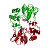
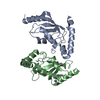
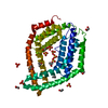
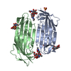
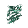
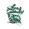
| Title | Crystal structure of an uncharacterized protein from Chlorobium tepidum | ||||||
 Components Components | uncharacterized protein | ||||||
 Keywords Keywords | structural genomics / unknown function / PSI-2 / Protein Structure Initiative / New York SGX Research Center for Structural Genomics / NYSGXRC | ||||||
| Function / homology |  Function and homology information Function and homology informationCHAD domain / CHAD domain / CHAD domain superfamily / CHAD / CHAD domain / CHAD domain / CHAD domain profile. / Alpha solenoid / Mainly Alpha Similarity search - Domain/homology | ||||||
| Biological species |  Chlorobaculum tepidum (bacteria) Chlorobaculum tepidum (bacteria) | ||||||
| Method |  X-RAY DIFFRACTION / SYNCHROTRON / SAD / Resolution: 2.09 Å X-RAY DIFFRACTION / SYNCHROTRON / SAD / Resolution: 2.09 Å | ||||||
 Authors Authors | Bonanno, J.B. / Dickey, M. / Bain, K.T. / Powell, A. / Ozyurt, S. / Smith, D. / Wasserman, S. / Sauder, J.M. / Burley, S.K. / Almo, S.C. / New York SGX Research Center for Structural Genomics (NYSGXRC) | ||||||
 Citation Citation | Journal: To be Published Title: Crystal structure of an uncharacterized protein from Chlorobium tepidum Authors: Bonanno, J.B. / Dickey, M. / Bain, K.T. / Powell, A. / Ozyurt, S. / Smith, D. / Wasserman, S. / Sauder, J.M. / Burley, S.K. / Almo, S.C. | ||||||
| History |
|
- Structure visualization
| Structure viewer | Molecule: MolmilJmol/JSmol |
|---|
- Downloads & links
-Download
| PDBx/mmCIF format | 3e0s.cif.gz | 130.1 KB | Display | PDBx/mmCIF format |
|---|---|---|---|---|
| PDB format | pdb3e0s.ent.gz | 100.9 KB | Display | PDB format |
| PDBx/mmJSON format | 3e0s.json.gz | Tree view | PDBx/mmJSON format | |
| Others |  Other downloads Other downloads |
-Validation report
| Arichive directory | https://data.pdbj.org/pub/pdb/validation_reports/e0/3e0sftp://data.pdbj.org/pub/pdb/validation_reports/e0/3e0s | HTTPS FTP |
|---|
-Related structure data
| Similar structure data | |
|---|---|
| Other databases |


-Links
 PDBj
PDBj- Assembly
| Deposited unit | 
| ||||||||
|---|---|---|---|---|---|---|---|---|---|
| 1 | 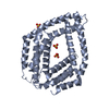
| ||||||||
| 2 | 
| ||||||||
| Unit cell |
| ||||||||
| Details | Authors state that the biological assembly is probably monomer |
-Components
| #1: Protein | Mass: 37352.508 Da / Num. of mol.: 2 / Fragment: residues 208-522 Source method: isolated from a genetically manipulated source Source: (gene. exp.) Chlorobaculum tepidum (bacteria) / Gene: CT0884 / Plasmid: modified pET26 / Production host: #2: Chemical | ChemComp-SO4 /   Mass: 96.063 Da / Num. of mol.: 5 / Source method: obtained synthetically / Formula: SO4 Mass: 96.063 Da / Num. of mol.: 5 / Source method: obtained synthetically / Formula: SO4#3: Water | ChemComp-HOH / |  Mass: 18.015 Da / Num. of mol.: 193 / Source method: isolated from a natural source / Formula: H2O Mass: 18.015 Da / Num. of mol.: 193 / Source method: isolated from a natural source / Formula: H2O |
|---|
-Experimental details
-Experiment
| Experiment | Method: X-RAY DIFFRACTION / Number of used crystals: 1 |
|---|
- Sample preparation
| Crystal | Density Matthews: 2.54 Å3/Da / Density % sol: 51.65 % |
|---|---|
| Crystal grow | Temperature: 294 K / Method: vapor diffusion / pH: 7 Details: 13% PEG 8K, 400mM lithium sulfate, pH 7.0, Vapor diffusion, temperature 294K |
-Data collection
| Diffraction | Mean temperature: 100 K |
|---|---|
| Diffraction source | Source: SYNCHROTRON / Site: APS  / Beamline: 31-ID / Wavelength: 0.97958 Å / Beamline: 31-ID / Wavelength: 0.97958 Å |
| Detector | Type: MAR CCD 165 mm / Detector: CCD / Date: Dec 8, 2007 |
| Radiation | Monochromator: diamond / Protocol: SINGLE WAVELENGTH / Monochromatic (M) / Laue (L): M / Scattering type: x-ray |
| Radiation wavelength | Wavelength: 0.97958 Å / Relative weight: 1 |
| Reflection | Resolution: 2.089→37.529 Å / Num. all: 43980 / Num. obs: 40989 / % possible obs: 93.2 % / Observed criterion σ(F): 0 / Observed criterion σ(I): 0 / Redundancy: 3.5 % / Biso Wilson estimate: 23.2 Å2 / Rmerge(I) obs: 0.089 / Rsym value: 0.089 / Net I/σ(I): 12.2 |
| Reflection shell | Resolution: 2.09→2.2 Å / Redundancy: 3.5 % / Rmerge(I) obs: 0.325 / Mean I/σ(I) obs: 3.9 / Num. measured all: 19090 / Num. unique all: 5467 / Rsym value: 0.325 / % possible all: 85.6 |
- Processing
| Software |
| ||||||||||||||||||||||||||||||||||||||||||||||||||||||||||||||||||||||||||||||||||||||||||
|---|---|---|---|---|---|---|---|---|---|---|---|---|---|---|---|---|---|---|---|---|---|---|---|---|---|---|---|---|---|---|---|---|---|---|---|---|---|---|---|---|---|---|---|---|---|---|---|---|---|---|---|---|---|---|---|---|---|---|---|---|---|---|---|---|---|---|---|---|---|---|---|---|---|---|---|---|---|---|---|---|---|---|---|---|---|---|---|---|---|---|---|
| Refinement | Method to determine structure: SAD / Resolution: 2.09→20 Å / Cor.coef. Fo:Fc: 0.921 / Cor.coef. Fo:Fc free: 0.877 / Occupancy max: 1 / Occupancy min: 0.5 / FOM work R set: 0.824 / SU B: 4.951 / SU ML: 0.137 / Cross valid method: THROUGHOUT / σ(F): 0 / σ(I): 0 / ESU R: 0.234 / ESU R Free: 0.208 / Stereochemistry target values: MAXIMUM LIKELIHOOD / Details: HYDROGENS HAVE BEEN ADDED IN THE RIDING POSITIONS
| ||||||||||||||||||||||||||||||||||||||||||||||||||||||||||||||||||||||||||||||||||||||||||
| Solvent computation | Ion probe radii: 0.8 Å / Shrinkage radii: 0.8 Å / VDW probe radii: 1.4 Å / Solvent model: BABINET MODEL WITH MASK | ||||||||||||||||||||||||||||||||||||||||||||||||||||||||||||||||||||||||||||||||||||||||||
| Displacement parameters | Biso max: 66.79 Å2 / Biso mean: 34.42 Å2 / Biso min: 12.41 Å2
| ||||||||||||||||||||||||||||||||||||||||||||||||||||||||||||||||||||||||||||||||||||||||||
| Refinement step | Cycle: LAST / Resolution: 2.09→20 Å
| ||||||||||||||||||||||||||||||||||||||||||||||||||||||||||||||||||||||||||||||||||||||||||
| Refine LS restraints |
| ||||||||||||||||||||||||||||||||||||||||||||||||||||||||||||||||||||||||||||||||||||||||||
| LS refinement shell | Resolution: 2.089→2.143 Å / Total num. of bins used: 20
|
