 Movie
Movie Controller


+ Open data

- Basic information
| Entry | Database: PDB / ID: 3dvo | ||||||
|---|---|---|---|---|---|---|---|
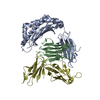
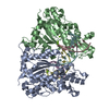
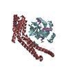
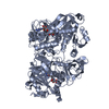
| Title | SgrAI with cognate DNA and calcium bound | ||||||
 Components Components |
| ||||||
 Keywords Keywords | HYDROLASE/DNA / restriction enzyme-DNA complex / HYDROLASE-DNA COMPLEX | ||||||
| Function / homology |  Function and homology information Function and homology information | ||||||
| Biological species |  Streptomyces griseus (bacteria) Streptomyces griseus (bacteria)synthetic construct (others) | ||||||
| Method |  X-RAY DIFFRACTION / SYNCHROTRON / MOLECULAR REPLACEMENT / Resolution: 1.892 Å X-RAY DIFFRACTION / SYNCHROTRON / MOLECULAR REPLACEMENT / Resolution: 1.892 Å | ||||||
 Authors Authors | Dunten, P.W. / Horton, N.C. / Little, E.J. | ||||||
 Citation Citation | Journal: Nucleic Acids Res. / Year: 2008 Title: The structure of SgrAI bound to DNA; recognition of an 8 base pair target. Authors: Dunten, P.W. / Little, E.J. / Gregory, M.T. / Manohar, V.M. / Dalton, M. / Hough, D. / Bitinaite, J. / Horton, N.C. | ||||||
| History |
|
- Structure visualization
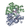
| Structure viewer | Molecule: MolmilJmol/JSmol |
|---|
- Downloads & links
-Download
| PDBx/mmCIF format | 3dvo.cif.gz | 325.3 KB | Display | PDBx/mmCIF format |
|---|---|---|---|---|
| PDB format | pdb3dvo.ent.gz | 257.3 KB | Display | PDB format |
| PDBx/mmJSON format | 3dvo.json.gz | Tree view | PDBx/mmJSON format | |
| Others |  Other downloads Other downloads |
-Validation report
| Summary document | 3dvo_validation.pdf.gz | 478.5 KB | Display | wwPDB validaton report |
|---|---|---|---|---|
| Full document | 3dvo_full_validation.pdf.gz | 495.9 KB | Display | |
| Data in XML | 3dvo_validation.xml.gz | 56.6 KB | Display | |
| Data in CIF | 3dvo_validation.cif.gz | 82.3 KB | Display | |
| Arichive directory | https://data.pdbj.org/pub/pdb/validation_reports/dv/3dvoftp://data.pdbj.org/pub/pdb/validation_reports/dv/3dvo | HTTPS FTP |
-Related structure data
| Related structure data | 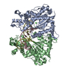 3dpgSC  3dw9C S: Starting model for refinement C: citing same article ( |
|---|---|
| Similar structure data |


-Links
 PDBj
PDBj


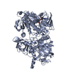
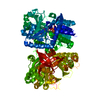
- Assembly
| Deposited unit | 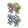
| ||||||||
|---|---|---|---|---|---|---|---|---|---|
| 1 | 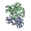
| ||||||||
| 2 | 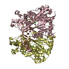
| ||||||||
| Unit cell |
|
-Components
| #1: DNA chain | Mass: 5517.567 Da / Num. of mol.: 4 / Source method: obtained synthetically / Details: includes the SgrAI recognition sequence / Source: (synth.) synthetic construct (others) #2: Protein | Mass: 37941.906 Da / Num. of mol.: 4 / Mutation: N63D Source method: isolated from a genetically manipulated source Details: coexpressed with MspI methyltransferase from pBAKMspIM Source: (gene. exp.) Streptomyces griseus (bacteria) / Gene: sgraIR / Plasmid: pET21a_SgrAIR / Production host: References: UniProt: Q9F6L0, type II site-specific deoxyribonuclease #3: Chemical | ChemComp-CA /   Mass: 40.078 Da / Num. of mol.: 8 / Source method: obtained synthetically / Formula: Ca Mass: 40.078 Da / Num. of mol.: 8 / Source method: obtained synthetically / Formula: Ca#4: Water | ChemComp-HOH / |  Mass: 18.015 Da / Num. of mol.: 776 / Source method: isolated from a natural source / Formula: H2O Mass: 18.015 Da / Num. of mol.: 776 / Source method: isolated from a natural source / Formula: H2O |
|---|
-Experimental details
-Experiment
| Experiment | Method: X-RAY DIFFRACTION / Number of used crystals: 1 |
|---|
- Sample preparation
| Crystal | Density Matthews: 2.5 Å3/Da / Density % sol: 50.78 % | ||||||||||||||||||||||||||||
|---|---|---|---|---|---|---|---|---|---|---|---|---|---|---|---|---|---|---|---|---|---|---|---|---|---|---|---|---|---|
| Crystal grow | Temperature: 290 K / Method: vapor diffusion, hanging drop Details: PEG4000, buffer, NaCl, CaCl2, VAPOR DIFFUSION, HANGING DROP, temperature 290K | ||||||||||||||||||||||||||||
| Components of the solutions |
|
-Data collection
| Diffraction source | Source: SYNCHROTRON / Site: SSRL  / Beamline: BL9-2 / Beamline: BL9-2 |
|---|---|
| Radiation | Protocol: SINGLE WAVELENGTH / Monochromatic (M) / Laue (L): M / Scattering type: x-ray |
| Radiation wavelength | Relative weight: 1 |
| Reflection | Highest resolution: 1.89 Å / Num. obs: 126080 / % possible obs: 92.8 % / Redundancy: 3.4 % / Rmerge(I) obs: 0.12 / Net I/σ(I): 6.5 |
- Processing
| Software | Name: PHENIX / Version: (phenix.refine) / Classification: refinement | ||||||||||||||||||||||||
|---|---|---|---|---|---|---|---|---|---|---|---|---|---|---|---|---|---|---|---|---|---|---|---|---|---|
| Refinement | Method to determine structure: MOLECULAR REPLACEMENT Starting model: pdb entry 3dpg Resolution: 1.892→41.068 Å / SU ML: 0.36 / Phase error: 28.53 / Stereochemistry target values: Engh & Huber
| ||||||||||||||||||||||||
| Solvent computation | Shrinkage radii: 0.9 Å / VDW probe radii: 1.11 Å / Solvent model: FLAT BULK SOLVENT MODEL / Bsol: 49.445 Å2 / ksol: 0.401 e/Å3 | ||||||||||||||||||||||||
| Refinement step | Cycle: LAST / Resolution: 1.892→41.068 Å
| ||||||||||||||||||||||||
| Refine LS restraints |
|
