 Movie
Movie Controller

[English] 日本語
 Yorodumi
- PDB-3dtf: Structural analysis of mycobacterial branched chain aminotransfer... -
+ Open data

- Basic information
| Entry | Database: PDB / ID: 3dtf | ||||||
|---|---|---|---|---|---|---|---|
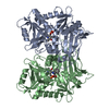
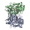
| Title | Structural analysis of mycobacterial branched chain aminotransferase- implications for inhibitor design | ||||||
 Components Components | Branched-chain amino acid aminotransferase | ||||||
 Keywords Keywords | TRANSFERASE / open twisted alpha/beta / Aminotransferase | ||||||
| Function / homology |  Function and homology information Function and homology informationprotein-pyridoxal-5-phosphate linkage via peptidyl-N6-pyridoxal phosphate-L-lysine / branched-chain amino acid metabolic process / branched-chain-amino-acid:2-oxoglutarate transaminase activity / L-valine:2-oxoglutarate transaminase activity / L-isoleucine:2-oxoglutarate transaminase activity / L-leucine:2-oxoglutarate transaminase activity / branched-chain-amino-acid transaminase / L-leucine biosynthetic process / L-valine biosynthetic process / : / pyridoxal phosphate binding Similarity search - Function | ||||||
| Biological species |  Mycobacterium smegmatis (bacteria) Mycobacterium smegmatis (bacteria) | ||||||
| Method |  X-RAY DIFFRACTION / SYNCHROTRON / MOLECULAR REPLACEMENT / Resolution: 2.2 Å X-RAY DIFFRACTION / SYNCHROTRON / MOLECULAR REPLACEMENT / Resolution: 2.2 Å | ||||||
 Authors Authors | Castell, A. / Unge, T. | ||||||
 Citation Citation | Journal: Acta Crystallogr.,Sect.D / Year: 2010 Title: Structural analysis of mycobacterial branched-chain aminotransferase: implications for inhibitor design. Authors: Castell, A. / Mille, C. / Unge, T. | ||||||
| History |
|
- Structure visualization
| Structure viewer | Molecule: MolmilJmol/JSmol |
|---|
- Downloads & links
-Download
| PDBx/mmCIF format | 3dtf.cif.gz | 155.3 KB | Display | PDBx/mmCIF format |
|---|---|---|---|---|
| PDB format | pdb3dtf.ent.gz | 121.5 KB | Display | PDB format |
| PDBx/mmJSON format | 3dtf.json.gz | Tree view | PDBx/mmJSON format | |
| Others |  Other downloads Other downloads |
-Validation report
| Arichive directory | https://data.pdbj.org/pub/pdb/validation_reports/dt/3dtfftp://data.pdbj.org/pub/pdb/validation_reports/dt/3dtf | HTTPS FTP |
|---|
-Related structure data
| Related structure data |  3jz6C 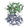 1ekfS C: citing same article ( S: Starting model for refinement |
|---|---|
| Similar structure data |


-Links
 PDBj
PDBj
- Assembly
| Deposited unit | 
| ||||||||
|---|---|---|---|---|---|---|---|---|---|
| 1 |
| ||||||||
| Unit cell |
|
-Components
| #1: Protein | Mass: 40366.215 Da / Num. of mol.: 2 Source method: isolated from a genetically manipulated source Source: (gene. exp.) Mycobacterium smegmatis (bacteria) / Gene: ilvE, MSMEG_4276 / Plasmid: pEXP5-CT/TOPO / Production host: References: UniProt: A0R066, branched-chain-amino-acid transaminase #2: Chemical |   Mass: 96.063 Da / Num. of mol.: 2 / Source method: obtained synthetically / Formula: SO4 Mass: 96.063 Da / Num. of mol.: 2 / Source method: obtained synthetically / Formula: SO4#3: Chemical |   Mass: 92.094 Da / Num. of mol.: 2 / Source method: obtained synthetically / Formula: C3H8O3 Mass: 92.094 Da / Num. of mol.: 2 / Source method: obtained synthetically / Formula: C3H8O3#4: Water | ChemComp-HOH / |  Mass: 18.015 Da / Num. of mol.: 342 / Source method: isolated from a natural source / Formula: H2O Mass: 18.015 Da / Num. of mol.: 342 / Source method: isolated from a natural source / Formula: H2O |
|---|
-Experimental details
-Experiment
| Experiment | Method: X-RAY DIFFRACTION / Number of used crystals: 1 |
|---|
- Sample preparation
| Crystal | Density Matthews: 2.3 Å3/Da / Density % sol: 46.57 % |
|---|---|
| Crystal grow | Temperature: 300 K / Method: vapor diffusion, sitting drop / pH: 5.5 Details: 0.4M Ammonium sulfate, 0.2M MES-HCl pH 5.5, 50% PEG 3350, VAPOR DIFFUSION, SITTING DROP, temperature 300K |
-Data collection
| Diffraction | Mean temperature: 100 K |
|---|---|
| Diffraction source | Source: SYNCHROTRON / Site: ESRF  / Beamline: ID14-2 / Wavelength: 0.933 Å / Beamline: ID14-2 / Wavelength: 0.933 Å |
| Detector | Type: ADSC QUANTUM 4 / Detector: CCD / Date: Oct 5, 2006 |
| Radiation | Protocol: SINGLE WAVELENGTH / Monochromatic (M) / Laue (L): M / Scattering type: x-ray |
| Radiation wavelength | Wavelength: 0.933 Å / Relative weight: 1 |
| Reflection | Resolution: 2.2→37.32 Å / Num. obs: 36343 / % possible obs: 98 % / Redundancy: 3.1 % / Biso Wilson estimate: 22.6 Å2 / Rmerge(I) obs: 0.072 / Rsym value: 0.086 / Net I/σ(I): 13 |
| Reflection shell | Resolution: 2.2→2.32 Å / Redundancy: 3.1 % / Rmerge(I) obs: 0.25 / Mean I/σ(I) obs: 4.9 / Num. unique all: 5342 / Rsym value: 0.3 / % possible all: 99.3 |
- Processing
| Software |
| |||||||||||||||||||||||||
|---|---|---|---|---|---|---|---|---|---|---|---|---|---|---|---|---|---|---|---|---|---|---|---|---|---|---|
| Refinement | Method to determine structure: MOLECULAR REPLACEMENT Starting model: PDB ENTRY 1EKF Resolution: 2.2→25 Å / Isotropic thermal model: anisotropic / Cross valid method: THROUGHOUT / Stereochemistry target values: Engh & Huber
| |||||||||||||||||||||||||
| Displacement parameters | Biso mean: 20.8 Å2 | |||||||||||||||||||||||||
| Refinement step | Cycle: LAST / Resolution: 2.2→25 Å
| |||||||||||||||||||||||||
| Refine LS restraints |
|
