 Movie
Movie Controller

[English] 日本語
 Yorodumi
- PDB-3dr5: Crystal structure of the Q8NRD3_CORGL protein from Corynebacteriu... -
+ Open data

- Basic information
| Entry | Database: PDB / ID: 3dr5 | ||||||
|---|---|---|---|---|---|---|---|
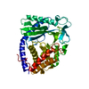
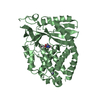
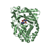
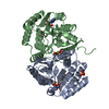
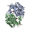
| Title | Crystal structure of the Q8NRD3_CORGL protein from Corynebacterium glutamicum. Northeast Structural Genomics Consortium target CgR117. | ||||||
 Components Components | Putative O-Methyltransferase | ||||||
 Keywords Keywords | structural genomics / unknown function / Q8NRD3 / Cgl1119 / PF01596 / O-METHYLTRANSFERASE / CgR117 / NESG / PSI-2 / Protein Structure Initiative / Northeast Structural Genomics Consortium / Methyltransferase / Transferase | ||||||
| Function / homology |  Function and homology information Function and homology information | ||||||
| Biological species |  Corynebacterium glutamicum (bacteria) Corynebacterium glutamicum (bacteria) | ||||||
| Method |  X-RAY DIFFRACTION / SYNCHROTRON / SAD / Resolution: 2.25 Å X-RAY DIFFRACTION / SYNCHROTRON / SAD / Resolution: 2.25 Å | ||||||
 Authors Authors | Vorobiev, S.M. / Su, M. / Seetharaman, J. / Wang, D. / Ciccosanti, C. / Foote, E.L. / Mao, L. / Xiao, R. / Acton, T.B. / Montelione, G.T. ...Vorobiev, S.M. / Su, M. / Seetharaman, J. / Wang, D. / Ciccosanti, C. / Foote, E.L. / Mao, L. / Xiao, R. / Acton, T.B. / Montelione, G.T. / Hunt, J.F. / Tong, L. / Northeast Structural Genomics Consortium (NESG) | ||||||
 Citation Citation | Journal: To be Published Title: Crystal structure of the Q8NRD3_CORGL protein from Corynebacterium glutamicum. Authors: Vorobiev, S.M. / Su, M. / Seetharaman, J. / Wang, D. / Ciccosanti, C. / Foote, E.L. / Mao, L. / Xiao, R. / Montelione, G.T. / Hunt, J.F. / Tong, L. | ||||||
| History |
|
- Structure visualization
| Structure viewer | Molecule: MolmilJmol/JSmol |
|---|
- Downloads & links
-Download
| PDBx/mmCIF format | 3dr5.cif.gz | 57.8 KB | Display | PDBx/mmCIF format |
|---|---|---|---|---|
| PDB format | pdb3dr5.ent.gz | 41.5 KB | Display | PDB format |
| PDBx/mmJSON format | 3dr5.json.gz | Tree view | PDBx/mmJSON format | |
| Others |  Other downloads Other downloads |
-Validation report
| Summary document | 3dr5_validation.pdf.gz | 432 KB | Display | wwPDB validaton report |
|---|---|---|---|---|
| Full document | 3dr5_full_validation.pdf.gz | 438.3 KB | Display | |
| Data in XML | 3dr5_validation.xml.gz | 13.8 KB | Display | |
| Data in CIF | 3dr5_validation.cif.gz | 18.6 KB | Display | |
| Arichive directory | https://data.pdbj.org/pub/pdb/validation_reports/dr/3dr5ftp://data.pdbj.org/pub/pdb/validation_reports/dr/3dr5 | HTTPS FTP |
-Related structure data
| Similar structure data | |
|---|---|
| Other databases |

-Links
 PDBj
PDBj- Assembly
| Deposited unit | 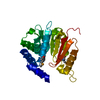
| ||||||||
|---|---|---|---|---|---|---|---|---|---|
| 1 | 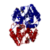
| ||||||||
| Unit cell |
| ||||||||
| Components on special symmetry positions |
| ||||||||
| Details | Authors state that the biological unit is Dimer according to aggregation screening. The second symmetry mate is generated by x,x-y+1,-z+7/6. |
-Components
| #1: Protein | Mass: 24005.309 Da / Num. of mol.: 1 Source method: isolated from a genetically manipulated source Source: (gene. exp.) Corynebacterium glutamicum (bacteria) / Strain: ATCC 13032/DSM 20300/JCM 1318/ LMG 3730/NCIMB 10025 / Gene: Cgl1119, cg1270 / Plasmid: pET21-23C / Production host: |
|---|---|
| #2: Water | ChemComp-HOH /  Mass: 18.015 Da / Num. of mol.: 170 / Source method: isolated from a natural source / Formula: H2O Mass: 18.015 Da / Num. of mol.: 170 / Source method: isolated from a natural source / Formula: H2O |
| Has protein modification | Y |
-Experimental details
-Experiment
| Experiment | Method: X-RAY DIFFRACTION / Number of used crystals: 1 |
|---|
- Sample preparation
| Crystal | Density Matthews: 2.33 Å3/Da / Density % sol: 47.13 % |
|---|---|
| Crystal grow | Temperature: 291 K / Method: vapor diffusion, hanging drop / pH: 6.15 Details: 10% PEG 4000, 0.1M magnesium chloride, 0.1M MES, pH6.15, VAPOR DIFFUSION, HANGING DROP, temperature 291K |
-Data collection
| Diffraction | Mean temperature: 100 K |
|---|---|
| Diffraction source | Source: SYNCHROTRON / Site: NSLS  / Beamline: X4A / Wavelength: 0.97945 Å / Beamline: X4A / Wavelength: 0.97945 Å |
| Detector | Type: ADSC QUANTUM 4 / Detector: CCD / Date: Jun 22, 2008 |
| Radiation | Protocol: SINGLE WAVELENGTH / Monochromatic (M) / Laue (L): M / Scattering type: x-ray |
| Radiation wavelength | Wavelength: 0.97945 Å / Relative weight: 1 |
| Reflection | Resolution: 2.25→50 Å / Num. obs: 19601 / % possible obs: 93.4 % / Observed criterion σ(F): 0 / Observed criterion σ(I): 0 / Redundancy: 20.1 % / Biso Wilson estimate: 22.9 Å2 / Rmerge(I) obs: 0.087 / Net I/σ(I): 22.3 |
| Reflection shell | Resolution: 2.25→2.33 Å / Redundancy: 19.4 % / Rmerge(I) obs: 0.283 / Mean I/σ(I) obs: 5.6 / Num. unique all: 8205 / % possible all: 69.2 |
- Processing
| Software |
| ||||||||||||||||||||||||||||||||||||||||||||||||||||||||||||
|---|---|---|---|---|---|---|---|---|---|---|---|---|---|---|---|---|---|---|---|---|---|---|---|---|---|---|---|---|---|---|---|---|---|---|---|---|---|---|---|---|---|---|---|---|---|---|---|---|---|---|---|---|---|---|---|---|---|---|---|---|---|
| Refinement | Method to determine structure: SAD / Resolution: 2.25→43.3 Å / Rfactor Rfree error: 0.008 / Data cutoff high absF: 281023.95 / Data cutoff low absF: 0 / Isotropic thermal model: RESTRAINED / Cross valid method: THROUGHOUT / σ(F): 2 / Stereochemistry target values: Engh & Huber
| ||||||||||||||||||||||||||||||||||||||||||||||||||||||||||||
| Solvent computation | Solvent model: FLAT MODEL / Bsol: 78.8008 Å2 / ksol: 0.335338 e/Å3 | ||||||||||||||||||||||||||||||||||||||||||||||||||||||||||||
| Displacement parameters | Biso mean: 42.6 Å2
| ||||||||||||||||||||||||||||||||||||||||||||||||||||||||||||
| Refine analyze |
| ||||||||||||||||||||||||||||||||||||||||||||||||||||||||||||
| Refinement step | Cycle: LAST / Resolution: 2.25→43.3 Å
| ||||||||||||||||||||||||||||||||||||||||||||||||||||||||||||
| Refine LS restraints |
| ||||||||||||||||||||||||||||||||||||||||||||||||||||||||||||
| LS refinement shell | Resolution: 2.25→2.39 Å / Rfactor Rfree error: 0.026 / Total num. of bins used: 6
| ||||||||||||||||||||||||||||||||||||||||||||||||||||||||||||
| Xplor file |
|
