 Movie
Movie Controller

[English] 日本語
 Yorodumi
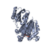
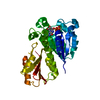
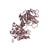
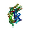
- PDB-3do8: The crystal structure of the protein with unknown function from A... -
+ Open data

- Basic information
| Entry | Database: PDB / ID: 3do8 | ||||||
|---|---|---|---|---|---|---|---|
| Title | The crystal structure of the protein with unknown function from Archaeoglobus fulgidus | ||||||
 Components Components | Phosphopantetheine adenylyltransferase | ||||||
 Keywords Keywords | TRANSFERASE / Protein with unknown function / Archaeoglobus fulgidus / Structural genomics / MCSG / PSI-2 / Protein Structure Initiative / Midwest Center for Structural Genomics / Coenzyme A biosynthesis / Cytoplasm / Nucleotidyltransferase | ||||||
| Function / homology |  Function and homology information Function and homology informationpantetheine-phosphate adenylyltransferase / pantetheine-phosphate adenylyltransferase activity / coenzyme A biosynthetic process / ATP binding / cytoplasm Similarity search - Function | ||||||
| Biological species |   Archaeoglobus fulgidus (archaea) Archaeoglobus fulgidus (archaea) | ||||||
| Method |  X-RAY DIFFRACTION / SYNCHROTRON / SAD / Resolution: 1.6 Å X-RAY DIFFRACTION / SYNCHROTRON / SAD / Resolution: 1.6 Å | ||||||
 Authors Authors | Zhang, R. / Wu, R. / Jedrzejczak, R. / Joachimiak, A. / Midwest Center for Structural Genomics (MCSG) | ||||||
 Citation Citation | Journal: To be Published / Year: 2008 Title: The crystal structure of the protein with unknown function from Archaeoglobus fulgidus Authors: Zhang, R. / Wu, R. / Jedrzejczak, R. / Joachimiak, A. | ||||||
| History |
|
- Structure visualization
| Structure viewer | Molecule: MolmilJmol/JSmol |
|---|
- Downloads & links
-Download
| PDBx/mmCIF format | 3do8.cif.gz | 121.8 KB | Display | PDBx/mmCIF format |
|---|---|---|---|---|
| PDB format | pdb3do8.ent.gz | 96.2 KB | Display | PDB format |
| PDBx/mmJSON format | 3do8.json.gz | Tree view | PDBx/mmJSON format | |
| Others |  Other downloads Other downloads |
-Validation report
| Arichive directory | https://data.pdbj.org/pub/pdb/validation_reports/do/3do8ftp://data.pdbj.org/pub/pdb/validation_reports/do/3do8 | HTTPS FTP |
|---|
-Related structure data
| Similar structure data | |
|---|---|
| Other databases |


-Links
 PDBj
PDBj- Assembly
| Deposited unit | 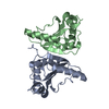
| ||||||||
|---|---|---|---|---|---|---|---|---|---|
| 1 |
| ||||||||
| Unit cell |
| ||||||||
| Details | This protein existed as dimer. The deposited Mol.A and Mol.B represent the dimer in the assymmetric unit. |
-Components
| #1: Protein | Mass: 16912.883 Da / Num. of mol.: 2 Source method: isolated from a genetically manipulated source Source: (gene. exp.) Archaeoglobus fulgidus (archaea) / Strain: DSM 4304 / Gene: coaD, AF_2206 / Plasmid: PDM68 / Production host:  References: UniProt: O28077, pantetheine-phosphate adenylyltransferase #2: Water | ChemComp-HOH / |  Mass: 18.015 Da / Num. of mol.: 216 / Source method: isolated from a natural source / Formula: H2O Mass: 18.015 Da / Num. of mol.: 216 / Source method: isolated from a natural source / Formula: H2O |
|---|
-Experimental details
-Experiment
| Experiment | Method: X-RAY DIFFRACTION / Number of used crystals: 1 |
|---|
- Sample preparation
| Crystal | Density Matthews: 2.12 Å3/Da / Density % sol: 42.08 % |
|---|---|
| Crystal grow | Temperature: 289 K / Method: vapor diffusion, sitting drop / pH: 7.5 Details: 0.1 M Hepes, pH7.5, 30%PEG MME, VAPOR DIFFUSION, SITTING DROP, temperature 289K |
-Data collection
| Diffraction | Mean temperature: 100 K |
|---|---|
| Diffraction source | Source: SYNCHROTRON / Site: APS  / Beamline: 19-BM / Wavelength: 0.9794 Å / Beamline: 19-BM / Wavelength: 0.9794 Å |
| Detector | Type: ADSC QUANTUM 315 / Detector: CCD / Date: Apr 7, 2008 / Details: mirrors |
| Radiation | Monochromator: Si 111 channel / Protocol: SINGLE WAVELENGTH / Monochromatic (M) / Laue (L): M / Scattering type: x-ray |
| Radiation wavelength | Wavelength: 0.9794 Å / Relative weight: 1 |
| Reflection | Resolution: 1.6→47.89 Å / Num. all: 35569 / Num. obs: 34228 / % possible obs: 96.23 % / Observed criterion σ(F): 2 / Observed criterion σ(I): 2 / Redundancy: 4.6 % / Biso Wilson estimate: 19 Å2 / Rmerge(I) obs: 0.059 / Net I/σ(I): 25.78 |
| Reflection shell | Resolution: 1.6→1.642 Å / Redundancy: 3.7 % / Rmerge(I) obs: 0.331 / Mean I/σ(I) obs: 2.25 / Num. unique all: 2757 / % possible all: 86.87 |
- Processing
| Software |
| |||||||||||||||||||||||||||||||||||||||||||||||||||||||||||||||||||||||||||||||||||||||||||||||||||||||||||||||||||||||||||||
|---|---|---|---|---|---|---|---|---|---|---|---|---|---|---|---|---|---|---|---|---|---|---|---|---|---|---|---|---|---|---|---|---|---|---|---|---|---|---|---|---|---|---|---|---|---|---|---|---|---|---|---|---|---|---|---|---|---|---|---|---|---|---|---|---|---|---|---|---|---|---|---|---|---|---|---|---|---|---|---|---|---|---|---|---|---|---|---|---|---|---|---|---|---|---|---|---|---|---|---|---|---|---|---|---|---|---|---|---|---|---|---|---|---|---|---|---|---|---|---|---|---|---|---|---|---|---|
| Refinement | Method to determine structure: SAD / Resolution: 1.6→47.89 Å / Cor.coef. Fo:Fc: 0.961 / Cor.coef. Fo:Fc free: 0.942 / SU B: 3.255 / SU ML: 0.053 / Cross valid method: THROUGHOUT / σ(F): 0 / σ(I): 0 / ESU R: 0.115 / ESU R Free: 0.092 Stereochemistry target values: MAXIMUM LIKELIHOOD WITH PHASES Details: HYDROGENS HAVE BEEN ADDED IN THE RIDING POSITIONS
| |||||||||||||||||||||||||||||||||||||||||||||||||||||||||||||||||||||||||||||||||||||||||||||||||||||||||||||||||||||||||||||
| Solvent computation | Ion probe radii: 0.8 Å / Shrinkage radii: 0.8 Å / VDW probe radii: 1.2 Å / Solvent model: MASK | |||||||||||||||||||||||||||||||||||||||||||||||||||||||||||||||||||||||||||||||||||||||||||||||||||||||||||||||||||||||||||||
| Displacement parameters | Biso mean: 18.06 Å2
| |||||||||||||||||||||||||||||||||||||||||||||||||||||||||||||||||||||||||||||||||||||||||||||||||||||||||||||||||||||||||||||
| Refinement step | Cycle: LAST / Resolution: 1.6→47.89 Å
| |||||||||||||||||||||||||||||||||||||||||||||||||||||||||||||||||||||||||||||||||||||||||||||||||||||||||||||||||||||||||||||
| Refine LS restraints |
| |||||||||||||||||||||||||||||||||||||||||||||||||||||||||||||||||||||||||||||||||||||||||||||||||||||||||||||||||||||||||||||
| LS refinement shell | Resolution: 1.6→1.642 Å / Total num. of bins used: 20
| |||||||||||||||||||||||||||||||||||||||||||||||||||||||||||||||||||||||||||||||||||||||||||||||||||||||||||||||||||||||||||||
| Refinement TLS params. | Method: refined / Origin x: 8.898 Å / Origin y: 12.035 Å / Origin z: 34.019 Å
| |||||||||||||||||||||||||||||||||||||||||||||||||||||||||||||||||||||||||||||||||||||||||||||||||||||||||||||||||||||||||||||
| Refinement TLS group |
|
