 Movie
Movie Controller

[English] 日本語
 Yorodumi
- PDB-3dez: Crystal structure of Orotate phosphoribosyltransferase from Strep... -
+ Open data

- Basic information
| Entry | Database: PDB / ID: 3dez | ||||||
|---|---|---|---|---|---|---|---|
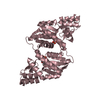
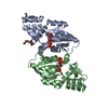
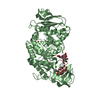
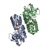
| Title | Crystal structure of Orotate phosphoribosyltransferase from Streptococcus mutans | ||||||
 Components Components | Orotate phosphoribosyltransferase | ||||||
 Keywords Keywords | TRANSFERASE / OROTATE PHOSPHORIBOSYLTRANSFERASE / Glycosyltransferase / Magnesium / Pyrimidine biosynthesis | ||||||
| Function / homology |  Function and homology information Function and homology informationorotate phosphoribosyltransferase / orotate phosphoribosyltransferase activity / pyrimidine nucleobase biosynthetic process / 'de novo' UMP biosynthetic process / magnesium ion binding Similarity search - Function | ||||||
| Biological species |  Streptococcus mutans (bacteria) Streptococcus mutans (bacteria) | ||||||
| Method |  X-RAY DIFFRACTION / SYNCHROTRON / MOLECULAR REPLACEMENT / Resolution: 2.4 Å X-RAY DIFFRACTION / SYNCHROTRON / MOLECULAR REPLACEMENT / Resolution: 2.4 Å | ||||||
 Authors Authors | Liu, C.P. / Gao, Z.Q. / Hou, H.F. / Li, L.F. / Su, X.D. / Dong, Y.H. | ||||||
 Citation Citation | Journal: Acta Crystallogr.,Sect.F / Year: 2010 Title: Structure of orotate phosphoribosyltransferase from the caries pathogen Streptococcus mutans Authors: Liu, C.P. / Xu, R. / Gao, Z.Q. / Xu, J.H. / Hou, H.F. / Li, L.Q. / She, Z. / Li, L.F. / Su, X.D. / Liu, P. / Dong, Y.H. | ||||||
| History |
|
- Structure visualization
| Structure viewer | Molecule: MolmilJmol/JSmol |
|---|
- Downloads & links
-Download
| PDBx/mmCIF format | 3dez.cif.gz | 91.6 KB | Display | PDBx/mmCIF format |
|---|---|---|---|---|
| PDB format | pdb3dez.ent.gz | 69.4 KB | Display | PDB format |
| PDBx/mmJSON format | 3dez.json.gz | Tree view | PDBx/mmJSON format | |
| Others |  Other downloads Other downloads |
-Validation report
| Arichive directory | https://data.pdbj.org/pub/pdb/validation_reports/de/3dezftp://data.pdbj.org/pub/pdb/validation_reports/de/3dez | HTTPS FTP |
|---|
-Related structure data
| Related structure data |  2aeeS S: Starting model for refinement |
|---|---|
| Similar structure data |


-Links
 PDBj
PDBj
- Assembly
| Deposited unit | 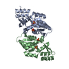
| ||||||||
|---|---|---|---|---|---|---|---|---|---|
| 1 |
| ||||||||
| Unit cell |
|
-Components
| #1: Protein | Mass: 26361.088 Da / Num. of mol.: 2 / Mutation: F92L Source method: isolated from a genetically manipulated source Source: (gene. exp.) Streptococcus mutans (bacteria) / Strain: UA159 / Gene: pyrE / Plasmid: pET28a / Production host: References: UniProt: Q8DTV2, orotate phosphoribosyltransferase #2: Chemical | ChemComp-SO4 /   Mass: 96.063 Da / Num. of mol.: 4 / Source method: obtained synthetically / Formula: SO4 Mass: 96.063 Da / Num. of mol.: 4 / Source method: obtained synthetically / Formula: SO4#3: Water | ChemComp-HOH / |  Mass: 18.015 Da / Num. of mol.: 89 / Source method: isolated from a natural source / Formula: H2O Mass: 18.015 Da / Num. of mol.: 89 / Source method: isolated from a natural source / Formula: H2O |
|---|
-Experimental details
-Experiment
| Experiment | Method: X-RAY DIFFRACTION / Number of used crystals: 1 |
|---|
- Sample preparation
| Crystal | Density Matthews: 2.39 Å3/Da / Density % sol: 48.45 % |
|---|---|
| Crystal grow | Temperature: 293 K / Method: vapor diffusion, hanging drop / pH: 8.6 Details: 0.1M Tris hydrochloride, 2.3M Ammonium sulfate, pH 8.6, VAPOR DIFFUSION, HANGING DROP, temperature 293K |
-Data collection
| Diffraction | Mean temperature: 100 K |
|---|---|
| Diffraction source | Source: SYNCHROTRON / Site: BSRF  / Beamline: 3W1A / Wavelength: 1 Å / Beamline: 3W1A / Wavelength: 1 Å |
| Detector | Type: MAR CCD 165 mm / Detector: CCD / Date: Mar 7, 2008 |
| Radiation | Protocol: SINGLE WAVELENGTH / Monochromatic (M) / Laue (L): M / Scattering type: x-ray |
| Radiation wavelength | Wavelength: 1 Å / Relative weight: 1 |
| Reflection | Resolution: 2.4→50 Å / Num. obs: 19803 / % possible obs: 85.7 % / Observed criterion σ(F): 1 / Observed criterion σ(I): 1 / Redundancy: 12 % |
| Reflection shell | Resolution: 2.4→2.49 Å / Redundancy: 10.9 % / Num. unique all: 2026 / % possible all: 100 |
- Processing
| Software |
| ||||||||||||||||
|---|---|---|---|---|---|---|---|---|---|---|---|---|---|---|---|---|---|
| Refinement | Method to determine structure: MOLECULAR REPLACEMENT Starting model: PDB ENTRY 2AEE Resolution: 2.4→50 Å / σ(F): 1 / Stereochemistry target values: maximum likelihood
| ||||||||||||||||
| Refinement step | Cycle: LAST / Resolution: 2.4→50 Å
| ||||||||||||||||
| Refine LS restraints |
|
