 Movie
Movie Controller


+ Open data

- Basic information
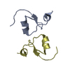
| Entry | Database: PDB / ID: 3cs5 | ||||||
|---|---|---|---|---|---|---|---|
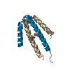
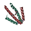
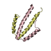
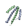
| Title | NblA protein from Synechococcus elongatus PCC 7942 | ||||||
 Components Components | Phycobilisome degradation protein nblA | ||||||
 Keywords Keywords | PHOTOSYNTHESIS / phycobilisome / nutrient stress / bleaching / helix-turn-helix / partial merohedral twinning | ||||||
| Function / homology | Phycobilisome degradation protein NblA / Phycobilisome degradation protein NblA / Phycobilisome degradation protein NblA superfamily / Phycobilisome degradation protein nblA / Helix Hairpins / Orthogonal Bundle / Mainly Alpha / Phycobilisome degradation protein NblA Function and homology information Function and homology information | ||||||
| Biological species |  Synechococcus sp. (bacteria) Synechococcus sp. (bacteria) | ||||||
| Method |  X-RAY DIFFRACTION / SYNCHROTRON / MOLECULAR REPLACEMENT / Resolution: 2.2 Å X-RAY DIFFRACTION / SYNCHROTRON / MOLECULAR REPLACEMENT / Resolution: 2.2 Å | ||||||
 Authors Authors | Dines, M. / Sendersky, E. / Schwarz, R. / Adir, N. | ||||||
 Citation Citation | Journal: J.Biol.Chem. / Year: 2008 Title: Structural, Functional, and Mutational Analysis of the NblA Protein Provides Insight into Possible Modes of Interaction with the Phycobilisome Authors: Dines, M. / Sendersky, E. / David, L. / Schwarz, R. / Adir, N. #1: Journal: J.Struct.Biol. / Year: 2007 Title: Crystallization of sparingly soluble stress-related proteins from cyanobacteria by controlled urea solublization Authors: Dines, M. / Sendersky, E. / Schwarz, R. / Adir, N. | ||||||
| History |
|
- Structure visualization
| Structure viewer | Molecule: MolmilJmol/JSmol |
|---|
- Downloads & links
-Download
| PDBx/mmCIF format | 3cs5.cif.gz | 51.6 KB | Display | PDBx/mmCIF format |
|---|---|---|---|---|
| PDB format | pdb3cs5.ent.gz | 38.7 KB | Display | PDB format |
| PDBx/mmJSON format | 3cs5.json.gz | Tree view | PDBx/mmJSON format | |
| Others |  Other downloads Other downloads |
-Validation report
| Arichive directory | https://data.pdbj.org/pub/pdb/validation_reports/cs/3cs5ftp://data.pdbj.org/pub/pdb/validation_reports/cs/3cs5 | HTTPS FTP |
|---|
-Related structure data
| Related structure data |  2q8vSC  2qdoC S: Starting model for refinement C: citing same article ( |
|---|---|
| Similar structure data |
-Links
 PDBj
PDBj- Assembly
| Deposited unit | 
| ||||||||
|---|---|---|---|---|---|---|---|---|---|
| 1 | 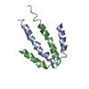
| ||||||||
| 2 | 
| ||||||||
| Unit cell |
| ||||||||
| Components on special symmetry positions |
|
-Components
| #1: Protein | Mass: 7060.076 Da / Num. of mol.: 4 Source method: isolated from a genetically manipulated source Source: (gene. exp.) Synechococcus sp. (bacteria) / Strain: PCC 7942 / Gene: nblA / Plasmid: pQE-70 / Production host: #2: Water | ChemComp-HOH / |  Mass: 18.015 Da / Num. of mol.: 27 / Source method: isolated from a natural source / Formula: H2O Mass: 18.015 Da / Num. of mol.: 27 / Source method: isolated from a natural source / Formula: H2O |
|---|
-Experimental details
-Experiment
| Experiment | Method: X-RAY DIFFRACTION / Number of used crystals: 1 |
|---|
- Sample preparation
| Crystal | Density Matthews: 3.81 Å3/Da / Density % sol: 67.75 % |
|---|---|
| Crystal grow | Temperature: 293 K / Method: vapor diffusion, hanging drop / pH: 8 Details: 25% ethylene glycol, pH8.0, VAPOR DIFFUSION, HANGING DROP, temperature 293.0K |
-Data collection
| Diffraction | Mean temperature: 100 K | ||||||||||||||||||
|---|---|---|---|---|---|---|---|---|---|---|---|---|---|---|---|---|---|---|---|
| Diffraction source | Source: SYNCHROTRON / Site: ESRF  / Beamline: ID14-1 / Wavelength: 0.934 Å / Beamline: ID14-1 / Wavelength: 0.934 Å | ||||||||||||||||||
| Detector | Type: ADSC QUANTUM 210 / Detector: CCD / Date: Mar 15, 2006 | ||||||||||||||||||
| Radiation | Protocol: SINGLE WAVELENGTH / Monochromatic (M) / Laue (L): M / Scattering type: x-ray | ||||||||||||||||||
| Radiation wavelength | Wavelength: 0.934 Å / Relative weight: 1 | ||||||||||||||||||
| Reflection twin |
| ||||||||||||||||||
| Reflection | Resolution: 2.2→20 Å / Num. obs: 21001 / % possible obs: 96.9 % / Redundancy: 6.8 % / Biso Wilson estimate: 20.1 Å2 / Rsym value: 0.057 / Net I/σ(I): 9.75 |
- Processing
| Software |
| ||||||||||||||||||||
|---|---|---|---|---|---|---|---|---|---|---|---|---|---|---|---|---|---|---|---|---|---|
| Refinement | Method to determine structure: MOLECULAR REPLACEMENT Starting model: PDB ENTRY 2Q8V Resolution: 2.2→20 Å / Isotropic thermal model: Isotropic / Cross valid method: THROUGHOUT / σ(F): 4 / Stereochemistry target values: Engh & Huber Details: This is a twinned structure, the detwin fraction is 0.479 and operator is 'h, -k, -l'.
| ||||||||||||||||||||
| Displacement parameters | Biso mean: 27.5 Å2 | ||||||||||||||||||||
| Refine analyze | Luzzati d res low obs: 5 Å / Luzzati sigma a obs: 0.509 Å | ||||||||||||||||||||
| Refinement step | Cycle: LAST / Resolution: 2.2→20 Å
| ||||||||||||||||||||
| Refine LS restraints |
|
