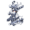
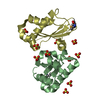
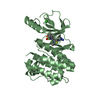
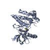
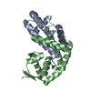
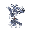
AUTHORS STATE THAT THE SIZE EXCLUSION CHROMATOGRAPHY WITH STATIC LIGHT SCATTERING SUPPORTS THE ASSIGNMENT OF A DIMER AS A SIGNIFICANT OLIGOMERIZATION STATE IN SOLUTION BUT CRYSTAL PACKING ANALYSIS DOES NOT REVEAL ANY INTERFACES THAT ARE LIKELY STABLE AS A DIMER.
-
Components
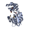
#1: Protein
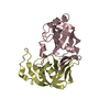
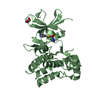
Cyclopropane-fatty-acyl-phospholipidsynthase
Mass: 31865.994 Da / Num. of mol.: 2 Source method: isolated from a genetically manipulated source Source: (gene. exp.) Anabaena variabilis ATCC 29413 (bacteria) Species: Anabaena variabilis / Strain: PCC 7937 / Gene: YP_321342.1, Ava_0823 / Plasmid: SpeedET / Production host: Escherichia coli (E. coli) / Strain (production host): HK100 References: UniProt: Q3MEY9, trans-aconitate 2-methyltransferase
Resolution: 1.9→29.921 Å / Num. obs: 40355 / % possible obs: 99.2 % / Observed criterion σ(I): -3 / Biso Wilson estimate: 27.041 Å2 / Rmerge(I) obs: 0.056 / Net I/σ(I): 9.39
Reflection shell
Resolution (Å)
Rmerge(I) obs
Mean I/σ(I) obs
Num. measured obs
Num. unique obs
Diffraction-ID
% possible all
1.9-1.97
0.502
1.7
14087
7587
1
95.6
1.97-2.05
0.386
2.2
14834
7775
1
99.8
2.05-2.14
0.301
2.9
13964
7330
1
99.8
2.14-2.25
0.231
3.8
14190
7469
1
99.6
2.25-2.39
0.173
5.1
14746
7678
1
99.8
2.39-2.58
0.125
6.5
15166
7887
1
99.7
2.58-2.84
0.084
9.3
14783
7635
1
99.8
2.84-3.25
0.051
14.4
14913
7640
1
99.8
3.25-4.08
0.033
21.3
14666
7553
1
99.6
4.08-29.921
0.023
26.5
15206
7711
1
99
-
Phasing
Phasing
Method: MAD
-
Processing
Software
Name
Version
Classification
NB
REFMAC
5.4.0067
refinement
PHENIX
refinement
SHELX
phasing
MolProbity
3beta29
modelbuilding
XSCALE
datascaling
PDB_EXTRACT
3
dataextraction
MAR345
CCD
datacollection
XDS
datareduction
SHELXD
phasing
autoSHARP
phasing
Refinement
Method to determine structure: MAD / Resolution: 1.9→29.921 Å / Cor.coef. Fo:Fc: 0.964 / Cor.coef. Fo:Fc free: 0.947 / SU B: 8.029 / SU ML: 0.119 / TLS residual ADP flag: LIKELY RESIDUAL / Cross valid method: THROUGHOUT / σ(F): 0 / ESU R: 0.154 / ESU R Free: 0.14 Stereochemistry target values: MAXIMUM LIKELIHOOD WITH PHASES Details: 1. HYDROGENS HAVE BEEN ADDED IN THE RIDING POSITIONS. 2. ATOM RECORDS CONTAIN RESIDUAL B FACTORS ONLY. 3. A MET-INHIBITION PROTOCOL WAS USED FOR SELENOMETHIONINE INCORPORATION DURING PROTEIN ...Details: 1. HYDROGENS HAVE BEEN ADDED IN THE RIDING POSITIONS. 2. ATOM RECORDS CONTAIN RESIDUAL B FACTORS ONLY. 3. A MET-INHIBITION PROTOCOL WAS USED FOR SELENOMETHIONINE INCORPORATION DURING PROTEIN EXPRESSION. THE OCCUPANCY OF THE SE ATOMS IN THE MSE RESIDUES WAS REDUCED TO 0.75 FOR THE REDUCED SCATTERING POWER DUE TO PARTIAL S-MET INCORPORATION. 4. BENZOIC ACID (BEZ) HAS BEEN MODELED BASED ON THE DENSITY. THIS ASSIGNMENT IS SUPPORTED BY A SALT LINK WITH ARG 251. HOWEVER, IT COULD BE SOME OTHER COMPOUND.
Rfactor
Num. reflection
% reflection
Selection details
Rfree
0.219
2024
5 %
RANDOM
Rwork
0.18
-
-
-
obs
0.182
40297
99.38 %
-
Solvent computation
Ion probe radii: 0.8 Å / Shrinkage radii: 0.8 Å / VDW probe radii: 1.2 Å / Solvent model: BABINET MODEL WITH MASK
In the structure databanks used in Yorodumi, some data are registered as the other names, "COVID-19 virus" and "2019-nCoV". Here are the details of the virus and the list of structure data.
Jan 31, 2019. EMDB accession codes are about to change! (news from PDBe EMDB page)
EMDB accession codes are about to change! (news from PDBe EMDB page)
The allocation of 4 digits for EMDB accession codes will soon come to an end. Whilst these codes will remain in use, new EMDB accession codes will include an additional digit and will expand incrementally as the available range of codes is exhausted. The current 4-digit format prefixed with “EMD-” (i.e. EMD-XXXX) will advance to a 5-digit format (i.e. EMD-XXXXX), and so on. It is currently estimated that the 4-digit codes will be depleted around Spring 2019, at which point the 5-digit format will come into force.
The EM Navigator/Yorodumi systems omit the EMD- prefix.
Related info.:Q: What is EMD? / ID/Accession-code notation in Yorodumi/EM Navigator
Yorodumi is a browser for structure data from EMDB, PDB, SASBDB, etc.
This page is also the successor to EM Navigator detail page, and also detail information page/front-end page for Omokage search.
The word "yorodu" (or yorozu) is an old Japanese word meaning "ten thousand". "mi" (miru) is to see.
Related info.:EMDB / PDB / SASBDB / Comparison of 3 databanks / Yorodumi Search / Aug 31, 2016. New EM Navigator & Yorodumi / Yorodumi Papers / Jmol/JSmol / Function and homology information / Changes in new EM Navigator and Yorodumi
 Movie
Movie Controller

 Yorodumi
 Open data
 Basic information
 Components
 Keywords
 Function and homology information
 Anabaena variabilis ATCC 29413 (bacteria)
 X-RAY DIFFRACTION /
  Authors
 Citation
 Structure visualization
 Downloads & links
 Other downloads

 PDBj
PDBj Assembly
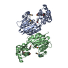


 Mass: 122.121 Da / Num. of mol.: 2 / Source method: obtained synthetically / Formula: C7H6O2
 Mass: 18.015 Da / Num. of mol.: 393 / Source method: isolated from a natural source / Formula: H2O
 Sample preparation
 / Beamline: BL9-2 / Wavelength: 0.91162, 0.97980, 0.97968
 Processing