 Movie
Movie Controller


+ Open data

- Basic information
| Entry | Database: PDB / ID: 3cba | ||||||
|---|---|---|---|---|---|---|---|
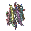
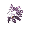
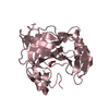
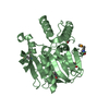
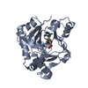
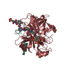
| Title | Crystal structure of Lipopeptide Detergent (LPD-12) (Hexagonal) | ||||||
 Components Components | LPD-12 | ||||||
 Keywords Keywords | DE NOVO PROTEIN / alpha helix / acyl chains / detergent / amphiphilic / lipopeptide / self-assembling peptide | ||||||
| Method |  X-RAY DIFFRACTION / SYNCHROTRON / MOLECULAR REPLACEMENT / Resolution: 1.7 Å X-RAY DIFFRACTION / SYNCHROTRON / MOLECULAR REPLACEMENT / Resolution: 1.7 Å | ||||||
 Authors Authors | Ho, D.N. / Pomroy, N.C. / Cuesta-Seijo, J.A. / Prive, G.G. | ||||||
 Citation Citation | Journal: To be Published Title: Packing and Twinning in Orthorhombic and Hexagonal Crystals of Lipopeptide Detergent (LPD-12) Authors: Ho, D.N. / Pomroy, N.C. / Cuesta-Seijo, J.A. / Prive, G.G. | ||||||
| History |
|
- Structure visualization
| Structure viewer | Molecule:  MolmilJmol/JSmol MolmilJmol/JSmol |
|---|
- Downloads & links
-Download
| PDBx/mmCIF format | 3cba.cif.gz | 79 KB | Display | PDBx/mmCIF format |
|---|---|---|---|---|
| PDB format | pdb3cba.ent.gz | 65 KB | Display | PDB format |
| PDBx/mmJSON format | 3cba.json.gz | Tree view | PDBx/mmJSON format | |
| Others |  Other downloads Other downloads |
-Validation report
| Arichive directory | https://data.pdbj.org/pub/pdb/validation_reports/cb/3cbaftp://data.pdbj.org/pub/pdb/validation_reports/cb/3cba | HTTPS FTP |
|---|
-Related structure data
| Related structure data | |
|---|---|
| Similar structure data |


-Links
 PDBj
PDBj
- Assembly
| Deposited unit | 
| ||||||||
|---|---|---|---|---|---|---|---|---|---|
| 1 | 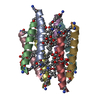
| ||||||||
| 2 | 
| ||||||||
| Unit cell |
|
-Components
| #1: Protein/peptide | Mass: 2826.394 Da / Num. of mol.: 12 / Source method: obtained synthetically / Details: de novo designed lipopeptide #2: Sugar | ChemComp-DMU /   Type: D-saccharide / Mass: 482.562 Da / Num. of mol.: 16 / Source method: obtained synthetically / Formula: C22H42O11 / Comment: detergent*YM Type: D-saccharide / Mass: 482.562 Da / Num. of mol.: 16 / Source method: obtained synthetically / Formula: C22H42O11 / Comment: detergent*YM#3: Water | ChemComp-HOH / |  Mass: 18.015 Da / Num. of mol.: 232 / Source method: isolated from a natural source / Formula: H2O Mass: 18.015 Da / Num. of mol.: 232 / Source method: isolated from a natural source / Formula: H2OHas protein modification | Y | |
|---|
-Experimental details
-Experiment
| Experiment | Method: X-RAY DIFFRACTION / Number of used crystals: 1 |
|---|
- Sample preparation
| Crystal | Density Matthews: 2.83 Å3/Da / Density % sol: 56.49 % |
|---|---|
| Crystal grow | Temperature: 295 K / Method: vapor diffusion, hanging drop / pH: 4.2 Details: 12% PEG 3350, 0.15M potassium phosphate, pH 4.2, VAPOR DIFFUSION, HANGING DROP, temperature 295K |
-Data collection
| Diffraction | Mean temperature: 100 K |
|---|---|
| Diffraction source | Source: SYNCHROTRON / Site: APS  / Beamline: 19-ID / Wavelength: 0.9793 Å / Beamline: 19-ID / Wavelength: 0.9793 Å |
| Detector | Type: ADSC QUANTUM 315 / Detector: CCD / Date: Dec 10, 2004 |
| Radiation | Monochromator: Rosenbaum-Rock high-resolution double-crystal monochromator. LN2 cooled first crystal, sagittal focusing 2nd crystal Protocol: SINGLE WAVELENGTH / Monochromatic (M) / Laue (L): M / Scattering type: x-ray |
| Radiation wavelength | Wavelength: 0.9793 Å / Relative weight: 1 |
| Reflection | Resolution: 1.7→50 Å / Num. all: 43769 / Num. obs: 43769 / % possible obs: 99.6 % / Observed criterion σ(F): 0 / Observed criterion σ(I): -3 / Redundancy: 5.9 % / Rsym value: 0.053 |
| Reflection shell | Resolution: 1.7→1.76 Å / Redundancy: 4.9 % / Rsym value: 0.26 / % possible all: 98.4 |
- Processing
| Software |
| ||||||||||||||||||||
|---|---|---|---|---|---|---|---|---|---|---|---|---|---|---|---|---|---|---|---|---|---|
| Refinement | Method to determine structure: MOLECULAR REPLACEMENT / Resolution: 1.7→10 Å / σ(F): 0 / Stereochemistry target values: Engh & Huber
| ||||||||||||||||||||
| Refinement step | Cycle: LAST / Resolution: 1.7→10 Å
| ||||||||||||||||||||
| Refine LS restraints |
|
