 Movie
Movie Controller

[English] 日本語
 Yorodumi
- PDB-3bvk: Structural basis for the iron uptake mechanism of Helicobacter py... -
+ Open data

- Basic information
| Entry | Database: PDB / ID: 3bvk | ||||||
|---|---|---|---|---|---|---|---|
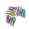
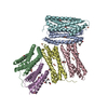
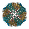
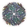
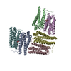
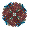
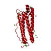
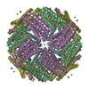
| Title | Structural basis for the iron uptake mechanism of Helicobacter pylori ferritin | ||||||
 Components Components | Ferritin | ||||||
 Keywords Keywords | OXIDOREDUCTASE / iron storage / Metal-binding | ||||||
| Function / homology |  Function and homology information Function and homology informationbacterial non-heme ferritin / ferroxidase activity / ferric iron binding / ferrous iron binding / iron ion transport / intracellular iron ion homeostasis / identical protein binding / cytosol Similarity search - Function | ||||||
| Biological species |   Helicobacter pylori (bacteria) Helicobacter pylori (bacteria) | ||||||
| Method |  X-RAY DIFFRACTION / SYNCHROTRON / MOLECULAR REPLACEMENT / Resolution: 1.5 Å X-RAY DIFFRACTION / SYNCHROTRON / MOLECULAR REPLACEMENT / Resolution: 1.5 Å | ||||||
 Authors Authors | Kim, K.H. / Cho, K.J. / Lee, J.H. / Shin, H.J. / Yang, I.S. | ||||||
 Citation Citation | Journal: To be Published Title: Structural basis for the iron uptake mechanism of Helicobacter pylori ferritin Authors: Kim, K.H. / Cho, K.J. / Lee, J.H. / Shin, H.J. / Yang, I.S. | ||||||
| History |
|
- Structure visualization
| Structure viewer | Molecule: MolmilJmol/JSmol |
|---|
- Downloads & links
-Download
| PDBx/mmCIF format | 3bvk.cif.gz | 450.1 KB | Display | PDBx/mmCIF format |
|---|---|---|---|---|
| PDB format | pdb3bvk.ent.gz | 369.4 KB | Display | PDB format |
| PDBx/mmJSON format | 3bvk.json.gz | Tree view | PDBx/mmJSON format | |
| Others |  Other downloads Other downloads |
-Validation report
| Summary document | 3bvk_validation.pdf.gz | 490.6 KB | Display | wwPDB validaton report |
|---|---|---|---|---|
| Full document | 3bvk_full_validation.pdf.gz | 500.7 KB | Display | |
| Data in XML | 3bvk_validation.xml.gz | 49.1 KB | Display | |
| Data in CIF | 3bvk_validation.cif.gz | 71.7 KB | Display | |
| Arichive directory | https://data.pdbj.org/pub/pdb/validation_reports/bv/3bvkftp://data.pdbj.org/pub/pdb/validation_reports/bv/3bvk | HTTPS FTP |
-Related structure data


-Links
 PDBj
PDBj


- Assembly
| Deposited unit | 
| ||||||||
|---|---|---|---|---|---|---|---|---|---|
| 1 | 
| ||||||||
| Unit cell |
| ||||||||
| Components on special symmetry positions |
|
-Components
| #1: Protein | Mass: 20957.465 Da / Num. of mol.: 6 Source method: isolated from a genetically manipulated source Source: (gene. exp.) Helicobacter pylori (bacteria) / Strain: J99 / Plasmid: pETDuet / Production host: #2: Chemical | ChemComp-FE /   Mass: 55.845 Da / Num. of mol.: 14 / Source method: obtained synthetically / Formula: Fe Mass: 55.845 Da / Num. of mol.: 14 / Source method: obtained synthetically / Formula: Fe#3: Chemical |   Mass: 92.094 Da / Num. of mol.: 3 / Source method: obtained synthetically / Formula: C3H8O3 Mass: 92.094 Da / Num. of mol.: 3 / Source method: obtained synthetically / Formula: C3H8O3#4: Water | ChemComp-HOH / |  Mass: 18.015 Da / Num. of mol.: 1053 / Source method: isolated from a natural source / Formula: H2O Mass: 18.015 Da / Num. of mol.: 1053 / Source method: isolated from a natural source / Formula: H2O |
|---|
-Experimental details
-Experiment
| Experiment | Method: X-RAY DIFFRACTION / Number of used crystals: 1 |
|---|
- Sample preparation
| Crystal | Density Matthews: 2.71 Å3/Da / Density % sol: 54.64 % |
|---|---|
| Crystal grow | Temperature: 295 K / Method: vapor diffusion, hanging drop / pH: 7.5 Details: 100 mM HEPES, 10% isopropanol, 200 mM sodium citrate, pH 7.5, VAPOR DIFFUSION, HANGING DROP, temperature 295K |
-Data collection
| Diffraction | Mean temperature: 100 K |
|---|---|
| Diffraction source | Source: SYNCHROTRON / Site: PAL/PLS  / Beamline: 4A / Wavelength: 1 Å / Beamline: 4A / Wavelength: 1 Å |
| Detector | Type: ADSC QUANTUM 210 / Detector: CCD / Date: Oct 19, 2006 |
| Radiation | Protocol: SINGLE WAVELENGTH / Monochromatic (M) / Laue (L): M / Scattering type: x-ray |
| Radiation wavelength | Wavelength: 1 Å / Relative weight: 1 |
| Reflection | Resolution: 1.5→50 Å / Num. all: 212629 / Num. obs: 208376 / % possible obs: 98 % / Rmerge(I) obs: 0.088 / Net I/σ(I): 31.3 |
| Reflection shell | Resolution: 1.5→1.55 Å / Rmerge(I) obs: 0.472 / Mean I/σ(I) obs: 2.9 / % possible all: 86.1 |
- Processing
| Software |
| |||||||||||||||||||||||||||||||||||||||||||||||||||||||||||||||||||||||||||||||||||||||||||||||||||||||||
|---|---|---|---|---|---|---|---|---|---|---|---|---|---|---|---|---|---|---|---|---|---|---|---|---|---|---|---|---|---|---|---|---|---|---|---|---|---|---|---|---|---|---|---|---|---|---|---|---|---|---|---|---|---|---|---|---|---|---|---|---|---|---|---|---|---|---|---|---|---|---|---|---|---|---|---|---|---|---|---|---|---|---|---|---|---|---|---|---|---|---|---|---|---|---|---|---|---|---|---|---|---|---|---|---|---|---|
| Refinement | Method to determine structure: MOLECULAR REPLACEMENT / Resolution: 1.5→23.61 Å / Cor.coef. Fo:Fc: 0.965 / Cor.coef. Fo:Fc free: 0.958 / SU B: 2.322 / SU ML: 0.04 / Cross valid method: THROUGHOUT / σ(F): 0 / ESU R: 0.081 / ESU R Free: 0.067 / Stereochemistry target values: MAXIMUM LIKELIHOOD / Details: HYDROGENS HAVE BEEN ADDED IN THE RIDING POSITIONS
| |||||||||||||||||||||||||||||||||||||||||||||||||||||||||||||||||||||||||||||||||||||||||||||||||||||||||
| Solvent computation | Ion probe radii: 0.8 Å / Shrinkage radii: 0.8 Å / VDW probe radii: 1.4 Å / Solvent model: MASK | |||||||||||||||||||||||||||||||||||||||||||||||||||||||||||||||||||||||||||||||||||||||||||||||||||||||||
| Displacement parameters | Biso mean: 16.681 Å2
| |||||||||||||||||||||||||||||||||||||||||||||||||||||||||||||||||||||||||||||||||||||||||||||||||||||||||
| Refinement step | Cycle: LAST / Resolution: 1.5→23.61 Å
| |||||||||||||||||||||||||||||||||||||||||||||||||||||||||||||||||||||||||||||||||||||||||||||||||||||||||
| Refine LS restraints |
| |||||||||||||||||||||||||||||||||||||||||||||||||||||||||||||||||||||||||||||||||||||||||||||||||||||||||
| LS refinement shell | Resolution: 1.501→1.54 Å / Total num. of bins used: 20
|
