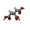
+ データを開く
- 基本情報
基本情報
| 登録情報 | データベース: PDB / ID: 3bsw | ||||||
|---|---|---|---|---|---|---|---|
| タイトル | PglD-citrate complex, from Campylobacter jejuni NCTC 11168 | ||||||
 要素 要素 | Acetyltransferase | ||||||
 キーワード キーワード | TRANSFERASE / left-hand beta helix / hexapeptide repeat / UDP / acetyl coenzyme Z / Rossmann fold / bacillosamine / campylobacter / pgl / N-linked glycosylation | ||||||
| 機能・相同性 |  機能・相同性情報 機能・相同性情報UDP-N-acetylbacillosamine N-acetyltransferase / protein N-linked glycosylation via asparagine / acyltransferase activity, transferring groups other than amino-acyl groups 類似検索 - 分子機能 | ||||||
| 生物種 |  | ||||||
| 手法 |  X線回折 / X線回折 /  シンクロトロン / シンクロトロン /  単一同系置換・異常分散 / 解像度: 1.77 Å 単一同系置換・異常分散 / 解像度: 1.77 Å | ||||||
| Model details | PglD with native substrate from Campylobacter jejuni, NCTC 11168 | ||||||
 データ登録者 データ登録者 | Olivier, N.B. / Imperiali, B. | ||||||
 引用 引用 |  ジャーナル: J.Biol.Chem. / 年: 2008 ジャーナル: J.Biol.Chem. / 年: 2008タイトル: Crystal structure and catalytic mechanism of PglD from Campylobacter jejuni. 著者: Olivier, N.B. / Imperiali, B. | ||||||
| 履歴 |
|
- 構造の表示
構造の表示
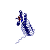
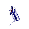
| 構造ビューア | 分子:  Molmil Molmil Jmol/JSmol Jmol/JSmol |
|---|
- ダウンロードとリンク
- ダウンロード
| PDBx/mmCIF形式 |  3bsw.cif.gz 3bsw.cif.gz | 54.7 KB | 表示 |  PDBx/mmCIF形式 PDBx/mmCIF形式 |
|---|---|---|---|---|
| PDB形式 |  pdb3bsw.ent.gz pdb3bsw.ent.gz | 38.3 KB | 表示 |  PDB形式 PDB形式 |
| PDBx/mmJSON形式 |  3bsw.json.gz 3bsw.json.gz | ツリー表示 |  PDBx/mmJSON形式 PDBx/mmJSON形式 | |
| その他 |  その他のダウンロード その他のダウンロード |
-検証レポート
| 文書・要旨 |  3bsw_validation.pdf.gz 3bsw_validation.pdf.gz | 440.2 KB | 表示 |  wwPDB検証レポート wwPDB検証レポート |
|---|---|---|---|---|
| 文書・詳細版 |  3bsw_full_validation.pdf.gz 3bsw_full_validation.pdf.gz | 440.9 KB | 表示 | |
| XML形式データ |  3bsw_validation.xml.gz 3bsw_validation.xml.gz | 11.7 KB | 表示 | |
| CIF形式データ |  3bsw_validation.cif.gz 3bsw_validation.cif.gz | 16.7 KB | 表示 | |
| アーカイブディレクトリ |  https://data.pdbj.org/pub/pdb/validation_reports/bs/3bsw https://data.pdbj.org/pub/pdb/validation_reports/bs/3bsw ftp://data.pdbj.org/pub/pdb/validation_reports/bs/3bsw ftp://data.pdbj.org/pub/pdb/validation_reports/bs/3bsw | HTTPS FTP |
-関連構造データ
- リンク
リンク
- 集合体
集合体
| 登録構造単位 | 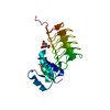
| ||||||||||||||||||
|---|---|---|---|---|---|---|---|---|---|---|---|---|---|---|---|---|---|---|---|
| 1 | 
| ||||||||||||||||||
| 単位格子 |
| ||||||||||||||||||
| Components on special symmetry positions |
| ||||||||||||||||||
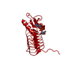
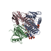
| 詳細 | biological unit is a trimer generated from the monomer asymmetric unit by the operations -x+y,-x,z and -y,x-y,z |
- 要素
要素
| #1: タンパク質 | 分子量: 21389.957 Da / 分子数: 1 / 由来タイプ: 組換発現 詳細: N-terminal residues GSA are non-native; resulted from removal of N-terminal His-tag by thrombin 由来: (組換発現)  株: NCTC 11168 / 遺伝子: pglD / プラスミド: pETGQ / 発現宿主:  参照: UniProt: Q0P9D1, UDP-N-acetylglucosamine diphosphorylase |
|---|---|
| #2: 化合物 | ChemComp-CIT / |
| #3: 水 | ChemComp-HOH / |
-実験情報
-実験
| 実験 | 手法:  X線回折 / 使用した結晶の数: 2 X線回折 / 使用した結晶の数: 2 |
|---|
- 試料調製
試料調製
| 結晶 |
| |||||||||||||||
|---|---|---|---|---|---|---|---|---|---|---|---|---|---|---|---|---|
| 結晶化 |
|
-データ収集
| 回折 |
| |||||||||||||||||||||||||||||||||||||||||||||||||||||||||||||||||||||||||||||
|---|---|---|---|---|---|---|---|---|---|---|---|---|---|---|---|---|---|---|---|---|---|---|---|---|---|---|---|---|---|---|---|---|---|---|---|---|---|---|---|---|---|---|---|---|---|---|---|---|---|---|---|---|---|---|---|---|---|---|---|---|---|---|---|---|---|---|---|---|---|---|---|---|---|---|---|---|---|---|
| 放射光源 | 由来:  シンクロトロン / サイト: シンクロトロン / サイト:  NSLS NSLS  / ビームライン: X6A / 波長: 0.9784 Å / ビームライン: X6A / 波長: 0.9784 Å | |||||||||||||||||||||||||||||||||||||||||||||||||||||||||||||||||||||||||||||
| 検出器 | タイプ: ADSC QUANTUM 210 / 検出器: CCD / 日付: 2007年2月7日 / 詳細: Toroidal focusing mirror | |||||||||||||||||||||||||||||||||||||||||||||||||||||||||||||||||||||||||||||
| 放射 | モノクロメーター: Si(111) channel cut monochromator / プロトコル: SINGLE WAVELENGTH / 単色(M)・ラウエ(L): M / 散乱光タイプ: x-ray | |||||||||||||||||||||||||||||||||||||||||||||||||||||||||||||||||||||||||||||
| 放射波長 | 波長: 0.9784 Å / 相対比: 1 | |||||||||||||||||||||||||||||||||||||||||||||||||||||||||||||||||||||||||||||
| Reflection | 冗長度: 5.6 % / Av σ(I) over netI: 5 / 数: 80527 / Rmerge(I) obs: 0.136 / Χ2: 1.01 / D res high: 2.2 Å / D res low: 30 Å / Num. obs: 14285 / % possible obs: 100 | |||||||||||||||||||||||||||||||||||||||||||||||||||||||||||||||||||||||||||||
| Diffraction reflection shell |
| |||||||||||||||||||||||||||||||||||||||||||||||||||||||||||||||||||||||||||||
| 反射 | 解像度: 1.77→30 Å / Num. all: 27293 / Num. obs: 27129 / % possible obs: 99.4 % / Observed criterion σ(F): 0 / Observed criterion σ(I): 0 / 冗長度: 5.7 % / Biso Wilson estimate: 25.11 Å2 / Rmerge(I) obs: 0.062 / Net I/σ(I): 27.7 | |||||||||||||||||||||||||||||||||||||||||||||||||||||||||||||||||||||||||||||
| 反射 シェル | 解像度: 1.77→1.83 Å / 冗長度: 5.7 % / Rmerge(I) obs: 0.571 / Mean I/σ(I) obs: 2.9 / Num. unique all: 2695 / % possible all: 99.6 |
-位相決定
| 位相決定 | 手法:  単一同系置換・異常分散 単一同系置換・異常分散 |
|---|
- 解析
解析
| ソフトウェア |
| ||||||||||||||||||||||||||||||||||||||||||||||||||||||||||||||||||||||||||||||||||||||||||
|---|---|---|---|---|---|---|---|---|---|---|---|---|---|---|---|---|---|---|---|---|---|---|---|---|---|---|---|---|---|---|---|---|---|---|---|---|---|---|---|---|---|---|---|---|---|---|---|---|---|---|---|---|---|---|---|---|---|---|---|---|---|---|---|---|---|---|---|---|---|---|---|---|---|---|---|---|---|---|---|---|---|---|---|---|---|---|---|---|---|---|---|
| 精密化 | 構造決定の手法:  単一同系置換・異常分散 / 解像度: 1.77→30 Å / Cor.coef. Fo:Fc: 0.962 / Cor.coef. Fo:Fc free: 0.961 / 交差検証法: THROUGHOUT / σ(F): 0 / σ(I): 0 / ESU R: 0.095 / ESU R Free: 0.09 / 立体化学のターゲット値: MAXIMUM LIKELIHOOD 単一同系置換・異常分散 / 解像度: 1.77→30 Å / Cor.coef. Fo:Fc: 0.962 / Cor.coef. Fo:Fc free: 0.961 / 交差検証法: THROUGHOUT / σ(F): 0 / σ(I): 0 / ESU R: 0.095 / ESU R Free: 0.09 / 立体化学のターゲット値: MAXIMUM LIKELIHOOD詳細: Heavy atom sites in the substructure were identified using SHELX-D with data collected at the Se peak wavelength and truncated to 2.5 . Three out of five possible selenium sites for a single ...詳細: Heavy atom sites in the substructure were identified using SHELX-D with data collected at the Se peak wavelength and truncated to 2.5 . Three out of five possible selenium sites for a single molecule of PglD in the asymmetric were located; CC All/weak=17.15/11.12, PATFOM 18.32. Structure factors from the native data were merged with initial phases using CAD (Riso=12%); phase extension to 1.77 and density modification were carried out using SHELX-E; values for contrast, connectivity, mean mapCC, and pseudo-free CC were 1.07, 0.96, 0.94, and 80.09%, respectively. The initial model was built with ARP/wARP (Perrakis et al., 2001) using the automated tracing function and manual adjustments were made using COOT (Emsley and Cowtan, 2004) and O (Jones et al., 1991).
| ||||||||||||||||||||||||||||||||||||||||||||||||||||||||||||||||||||||||||||||||||||||||||
| 溶媒の処理 | イオンプローブ半径: 0.8 Å / 減衰半径: 0.8 Å / VDWプローブ半径: 1.2 Å / 溶媒モデル: MASK | ||||||||||||||||||||||||||||||||||||||||||||||||||||||||||||||||||||||||||||||||||||||||||
| 原子変位パラメータ | Biso mean: 25.892 Å2
| ||||||||||||||||||||||||||||||||||||||||||||||||||||||||||||||||||||||||||||||||||||||||||
| 精密化ステップ | サイクル: LAST / 解像度: 1.77→30 Å
| ||||||||||||||||||||||||||||||||||||||||||||||||||||||||||||||||||||||||||||||||||||||||||
| 拘束条件 |
| ||||||||||||||||||||||||||||||||||||||||||||||||||||||||||||||||||||||||||||||||||||||||||
| LS精密化 シェル | 解像度: 1.77→1.816 Å / Total num. of bins used: 20
|
 ムービー
ムービー コントローラー






 PDBj
PDBj


