 Movie
Movie Controller

[English] 日本語
 Yorodumi
- PDB-3bk6: Crystal structure of a core domain of stomatin from Pyrococcus ho... -
+ Open data

- Basic information
| Entry | Database: PDB / ID: 3bk6 | ||||||
|---|---|---|---|---|---|---|---|
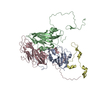
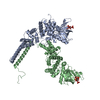
| Title | Crystal structure of a core domain of stomatin from Pyrococcus horikoshii | ||||||
 Components Components | PH stomatin | ||||||
 Keywords Keywords | MEMBRANE PROTEIN / stomatin / archaea / Pyrococcus horikoshii / trimer / coiled-coil / flotillin / SPFH / membrane fusion / trafficking / Transmembrane | ||||||
| Function / homology |  Function and homology information Function and homology information | ||||||
| Biological species |   Pyrococcus horikoshii (archaea) Pyrococcus horikoshii (archaea) | ||||||
| Method |  X-RAY DIFFRACTION / SYNCHROTRON / MAD / Resolution: 3.2 Å X-RAY DIFFRACTION / SYNCHROTRON / MAD / Resolution: 3.2 Å | ||||||
 Authors Authors | Yokoyama, H. / Fujii, S. / Matsui, I. | ||||||
 Citation Citation | Journal: J.Mol.Biol. / Year: 2008 Title: Crystal structure of a core domain of stomatin from Pyrococcus horikoshii Illustrates a novel trimeric and coiled-coil fold Authors: Yokoyama, H. / Fujii, S. / Matsui, I. #1: Journal: J.Mol.Biol. / Year: 2006Title: Molecular structure of a novel membrane protease specific for a stomatin homolog from the hyperthermophilic archaeon Pyrococcus horikoshii Authors: Yokoyama, H. / Matsui, E. / Akiba, T. / Harata, K. / Matsui, I. | ||||||
| History |
|
- Structure visualization
| Structure viewer | Molecule: MolmilJmol/JSmol |
|---|
- Downloads & links
-Download
| PDBx/mmCIF format | 3bk6.cif.gz | 102.4 KB | Display | PDBx/mmCIF format |
|---|---|---|---|---|
| PDB format | pdb3bk6.ent.gz | 80.4 KB | Display | PDB format |
| PDBx/mmJSON format | 3bk6.json.gz | Tree view | PDBx/mmJSON format | |
| Others |  Other downloads Other downloads |
-Validation report
| Arichive directory | https://data.pdbj.org/pub/pdb/validation_reports/bk/3bk6ftp://data.pdbj.org/pub/pdb/validation_reports/bk/3bk6 | HTTPS FTP |
|---|
-Related structure data
| Similar structure data |
|---|


-Links
 PDBj
PDBj
- Assembly
| Deposited unit | 
| ||||||||
|---|---|---|---|---|---|---|---|---|---|
| 1 |
| ||||||||
| Unit cell |
|
-Components
| #1: Protein | Mass: 21297.398 Da / Num. of mol.: 3 / Fragment: Residues 56-234 Source method: isolated from a genetically manipulated source Source: (gene. exp.) Pyrococcus horikoshii (archaea) / Gene: PH1511 / Plasmid: pET21b / Production host:  |
|---|
-Experimental details
-Experiment
| Experiment | Method: X-RAY DIFFRACTION / Number of used crystals: 2 |
|---|
- Sample preparation
| Crystal | Density Matthews: 4.27 Å3/Da / Density % sol: 71.19 % |
|---|---|
| Crystal grow | Temperature: 293 K / Method: vapor diffusion, hanging drop / pH: 7.5 Details: 20% MPD, 0.2M ammonium sulfate, 0.1M HEPES-NaOH, 6% 1,5-diaminopentane-HCl, pH 7.5, VAPOR DIFFUSION, HANGING DROP, temperature 293.0K |
-Data collection
| Diffraction | Mean temperature: 100 K | |||||||||||||||
|---|---|---|---|---|---|---|---|---|---|---|---|---|---|---|---|---|
| Diffraction source | Source: SYNCHROTRON / Site: Photon Factory  / Beamline: BL-6A / Wavelength: 0.9791, 0.9796, 0.9900, 1.0000 / Beamline: BL-6A / Wavelength: 0.9791, 0.9796, 0.9900, 1.0000 | |||||||||||||||
| Detector | Type: ADSC QUAMTUM 4r / Detector: CCD / Date: Nov 24, 2005 / Details: mirror | |||||||||||||||
| Radiation | Monochromator: Si(111) / Protocol: MAD / Monochromatic (M) / Laue (L): M / Scattering type: x-ray | |||||||||||||||
| Radiation wavelength |
| |||||||||||||||
| Reflection | Resolution: 3.2→50 Å / Num. obs: 17735 / % possible obs: 99.9 % / Redundancy: 3.8 % / Biso Wilson estimate: 114.6 Å2 / Rmerge(I) obs: 0.058 / Net I/σ(I): 33.5 | |||||||||||||||
| Reflection shell | Resolution: 3.2→3.31 Å / Rmerge(I) obs: 0.474 / Mean I/σ(I) obs: 3.6 / % possible all: 100 |
- Processing
| Software |
| ||||||||||||||||||||||||||||||||||||||||||||||||||||||||||||||||||||||||||||||||||||||||||
|---|---|---|---|---|---|---|---|---|---|---|---|---|---|---|---|---|---|---|---|---|---|---|---|---|---|---|---|---|---|---|---|---|---|---|---|---|---|---|---|---|---|---|---|---|---|---|---|---|---|---|---|---|---|---|---|---|---|---|---|---|---|---|---|---|---|---|---|---|---|---|---|---|---|---|---|---|---|---|---|---|---|---|---|---|---|---|---|---|---|---|---|
| Refinement | Method to determine structure: MAD / Resolution: 3.2→19.97 Å / Cor.coef. Fo:Fc: 0.954 / Cor.coef. Fo:Fc free: 0.917 / SU B: 18.023 / SU ML: 0.316 / Cross valid method: THROUGHOUT / ESU R: 1.343 / ESU R Free: 0.423 / Stereochemistry target values: MAXIMUM LIKELIHOOD
| ||||||||||||||||||||||||||||||||||||||||||||||||||||||||||||||||||||||||||||||||||||||||||
| Solvent computation | Ion probe radii: 0.8 Å / Shrinkage radii: 0.8 Å / VDW probe radii: 1.4 Å / Solvent model: MASK | ||||||||||||||||||||||||||||||||||||||||||||||||||||||||||||||||||||||||||||||||||||||||||
| Displacement parameters | Biso mean: 101.114 Å2 | ||||||||||||||||||||||||||||||||||||||||||||||||||||||||||||||||||||||||||||||||||||||||||
| Refine analyze |
| ||||||||||||||||||||||||||||||||||||||||||||||||||||||||||||||||||||||||||||||||||||||||||
| Refinement step | Cycle: LAST / Resolution: 3.2→19.97 Å
| ||||||||||||||||||||||||||||||||||||||||||||||||||||||||||||||||||||||||||||||||||||||||||
| Refine LS restraints |
| ||||||||||||||||||||||||||||||||||||||||||||||||||||||||||||||||||||||||||||||||||||||||||
| LS refinement shell | Resolution: 3.201→3.282 Å / Total num. of bins used: 20
|
