 Movie
Movie Controller

[English] 日本語
 Yorodumi
- PDB-3b4t: Crystal structure of Mycobacterium tuberculosis RNase PH, the Myc... -
+ Open data

- Basic information
| Entry | Database: PDB / ID: 3b4t | ||||||
|---|---|---|---|---|---|---|---|
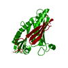
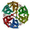
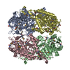
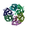
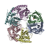
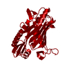
| Title | Crystal structure of Mycobacterium tuberculosis RNase PH, the Mycobacterium tuberculosis Structural Genomics Consortium target Rv1340 | ||||||
 Components Components | Ribonuclease PH | ||||||
 Keywords Keywords | TRANSFERASE / RNase / tRNA nucleotidyltransferase / ribonuclease / RPHA / Structural Genomics / TBSGC / Mycobacterium tuberculosis Structural Genomics Consortium / tRNA processing / TB Structural Genomics Consortium / PSI / Protein Structure Initiative | ||||||
| Function / homology |  Function and homology information Function and homology informationtRNA nucleotidyltransferase / tRNA nucleotidyltransferase activity / rRNA 3'-end processing / rRNA catabolic process / tRNA processing / rRNA processing / 3'-5'-RNA exonuclease activity / tRNA binding / RNA binding / plasma membrane Similarity search - Function | ||||||
| Biological species |   Mycobacterium tuberculosis (bacteria) Mycobacterium tuberculosis (bacteria) | ||||||
| Method |  X-RAY DIFFRACTION / SYNCHROTRON / MOLECULAR REPLACEMENT / Resolution: 2.1 Å X-RAY DIFFRACTION / SYNCHROTRON / MOLECULAR REPLACEMENT / Resolution: 2.1 Å | ||||||
 Authors Authors | Antczak, A.J. / Berger, J.M. / Lekin, T. / Segelke, B.W. / Mycobacterium Tuberculosis Structural Proteomics Project (XMTB) / TB Structural Genomics Consortium (TBSGC) | ||||||
 Citation Citation | Journal: To be Published Title: 2.1 A Crystal structure of RNase PH from Mycobacterium tuberculosis. Authors: Antczak, A.J. / Lekin, T. / Segelke, B.W. / Berger, J.M. | ||||||
| History |
|
- Structure visualization
| Structure viewer | Molecule: MolmilJmol/JSmol |
|---|
- Downloads & links
-Download
| PDBx/mmCIF format | 3b4t.cif.gz | 295.3 KB | Display | PDBx/mmCIF format |
|---|---|---|---|---|
| PDB format | pdb3b4t.ent.gz | 240.1 KB | Display | PDB format |
| PDBx/mmJSON format | 3b4t.json.gz | Tree view | PDBx/mmJSON format | |
| Others |  Other downloads Other downloads |
-Validation report
| Arichive directory | https://data.pdbj.org/pub/pdb/validation_reports/b4/3b4tftp://data.pdbj.org/pub/pdb/validation_reports/b4/3b4t | HTTPS FTP |
|---|
-Related structure data
| Related structure data |  1r6mS S: Starting model for refinement |
|---|---|
| Similar structure data | |
| Other databases |


-Links
 PDBj
PDBj
- Assembly
| Deposited unit | 
| ||||||||
|---|---|---|---|---|---|---|---|---|---|
| 1 |
| ||||||||
| Unit cell |
|
-Components
| #1: Protein | Mass: 27631.283 Da / Num. of mol.: 6 Source method: isolated from a genetically manipulated source Source: (gene. exp.) Mycobacterium tuberculosis (bacteria) / Strain: H37Rv / Gene: rph, rphA, Rv1340, MT1381, MTCY130.25, MTCY02B10.04 / Plasmid: pET22b / Production host: References: UniProt: Q10628, UniProt: P9WGZ7*PLUS, tRNA nucleotidyltransferase #2: Chemical | ChemComp-PO4 /   Mass: 94.971 Da / Num. of mol.: 10 / Source method: obtained synthetically / Formula: PO4 Mass: 94.971 Da / Num. of mol.: 10 / Source method: obtained synthetically / Formula: PO4#3: Water | ChemComp-HOH / |  Mass: 18.015 Da / Num. of mol.: 1107 / Source method: isolated from a natural source / Formula: H2O Mass: 18.015 Da / Num. of mol.: 1107 / Source method: isolated from a natural source / Formula: H2OHas protein modification | Y | |
|---|
-Experimental details
-Experiment
| Experiment | Method: X-RAY DIFFRACTION / Number of used crystals: 1 |
|---|
- Sample preparation
| Crystal | Density Matthews: 2.66 Å3/Da / Density % sol: 53.78 % Description: THE STRUCTURE FACTOR FILE CONTAINS FRIEDEL PAIRS |
|---|---|
| Crystal grow | Temperature: 298 K / pH: 7.5 Details: 22.5% PEG 2000, 0.1M MOPS pH 7.5, 0.085M NaH2PO4, 0.085M K2HPO4, VAPOR DIFFUSION, SITTING DROP, temperature 298.0K, pH 7.50 |
-Data collection
| Diffraction | Mean temperature: 100 K |
|---|---|
| Diffraction source | Source: SYNCHROTRON / Site: ALS  / Beamline: 8.3.1 / Wavelength: 1.11587 / Beamline: 8.3.1 / Wavelength: 1.11587 |
| Detector | Type: ADSC QUANTUM 315 / Detector: CCD / Date: Sep 2, 2005 |
| Radiation | Monochromator: DOUBLE FLAT CRYSTAL, SI(111) / Protocol: SINGLE WAVELENGTH / Monochromatic (M) / Laue (L): M / Scattering type: x-ray |
| Radiation wavelength | Wavelength: 1.11587 Å / Relative weight: 1 |
| Reflection | Resolution: 2.09→78.811 Å / Num. obs: 399198 / % possible obs: 100 % / Observed criterion σ(I): 1 / Redundancy: 3.8 % / Biso Wilson estimate: 28 Å2 / Rmerge(I) obs: 0.036 / Rsym value: 0.054 / Net I/σ(I): 22.2 |
| Reflection shell | Resolution: 2.09→2.18 Å / Redundancy: 3.7 % / Rmerge(I) obs: 0.422 / Mean I/σ(I) obs: 2.58 / Rsym value: 0.54 / % possible all: 99.7 |
- Processing
| Software |
| |||||||||||||||||||||||||
|---|---|---|---|---|---|---|---|---|---|---|---|---|---|---|---|---|---|---|---|---|---|---|---|---|---|---|
| Refinement | Method to determine structure: MOLECULAR REPLACEMENT Starting model: PDB ENTRY 1R6M Resolution: 2.1→39.69 Å / Isotropic thermal model: ANISOTROPIC / σ(F): 0 / Stereochemistry target values: Engh & Huber / Details: THE FRIEDEL PAIRS WERE USED IN PHASING
| |||||||||||||||||||||||||
| Displacement parameters | Biso mean: 39.67 Å2
| |||||||||||||||||||||||||
| Refinement step | Cycle: LAST / Resolution: 2.1→39.69 Å
| |||||||||||||||||||||||||
| LS refinement shell | Resolution: 2.1→2.12 Å
|
