 Movie
Movie Controller

[English] 日本語
 Yorodumi
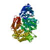
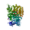
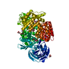
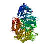
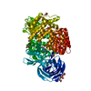
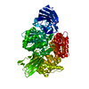
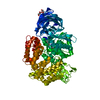
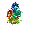
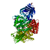
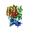
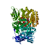
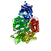
- PDB-3b37: Crystal structure of E. coli Aminopeptidase N in complex with Tyrosine -
+ Open data

- Basic information
| Entry | Database: PDB / ID: 3b37 | ||||||
|---|---|---|---|---|---|---|---|
| Title | Crystal structure of E. coli Aminopeptidase N in complex with Tyrosine | ||||||
 Components Components | Aminopeptidase N | ||||||
 Keywords Keywords | HYDROLASE / Aminopeptidase N / protease / thermolysin / tyrosine / Membrane / Metal-binding / Metalloprotease | ||||||
| Function / homology |  Function and homology information Function and homology informationmembrane alanyl aminopeptidase / alanyl aminopeptidase activity / aminopeptidase activity / metallopeptidase activity / proteolysis / zinc ion binding / identical protein binding / plasma membrane Similarity search - Function | ||||||
| Biological species |  | ||||||
| Method |  X-RAY DIFFRACTION / SYNCHROTRON / Resolution: 1.7 Å X-RAY DIFFRACTION / SYNCHROTRON / Resolution: 1.7 Å | ||||||
 Authors Authors | Addlagatta, A. | ||||||
 Citation Citation | Journal: Biochemistry / Year: 2008 Title: Structural basis for the unusual specificity of Escherichia coli aminopeptidase N. Authors: Addlagatta, A. / Gay, L. / Matthews, B.W. | ||||||
| History |
|
- Structure visualization
| Structure viewer | Molecule: MolmilJmol/JSmol |
|---|
- Downloads & links
-Download
| PDBx/mmCIF format | 3b37.cif.gz | 208.2 KB | Display | PDBx/mmCIF format |
|---|---|---|---|---|
| PDB format | pdb3b37.ent.gz | 161.6 KB | Display | PDB format |
| PDBx/mmJSON format | 3b37.json.gz | Tree view | PDBx/mmJSON format | |
| Others |  Other downloads Other downloads |
-Validation report
| Arichive directory | https://data.pdbj.org/pub/pdb/validation_reports/b3/3b37ftp://data.pdbj.org/pub/pdb/validation_reports/b3/3b37 | HTTPS FTP |
|---|
-Related structure data
| Related structure data |  3b2pC 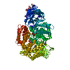 3b2xC 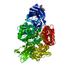 3b34C  3b3bC  2hpoS C: citing same article ( S: Starting model for refinement |
|---|---|
| Similar structure data |


-Links
 PDBj
PDBj

- Assembly
| Deposited unit | 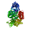
| ||||||||
|---|---|---|---|---|---|---|---|---|---|
| 1 |
| ||||||||
| Unit cell |
|
-Components
-Protein , 1 types, 1 molecules A
| #1: Protein | Mass: 101441.195 Da / Num. of mol.: 1 Source method: isolated from a genetically manipulated source Source: (gene. exp.) |
|---|
-Non-polymers , 6 types, 761 molecules 


| #2: Chemical | ChemComp-ZN /  Mass: 65.409 Da / Num. of mol.: 1 / Source method: obtained synthetically / Formula: Zn Mass: 65.409 Da / Num. of mol.: 1 / Source method: obtained synthetically / Formula: Zn | ||||||||
|---|---|---|---|---|---|---|---|---|---|
| #3: Chemical |  Mass: 22.990 Da / Num. of mol.: 2 / Source method: obtained synthetically / Formula: Na Mass: 22.990 Da / Num. of mol.: 2 / Source method: obtained synthetically / Formula: Na#4: Chemical | ChemComp-TYR / |  Type: L-peptide linking / Mass: 181.189 Da / Num. of mol.: 1 / Source method: obtained synthetically / Formula: C9H11NO3 Type: L-peptide linking / Mass: 181.189 Da / Num. of mol.: 1 / Source method: obtained synthetically / Formula: C9H11NO3#5: Chemical | ChemComp-MLI / |  Mass: 102.046 Da / Num. of mol.: 1 / Source method: obtained synthetically / Formula: C3H2O4 Mass: 102.046 Da / Num. of mol.: 1 / Source method: obtained synthetically / Formula: C3H2O4#6: Chemical | ChemComp-GOL /  Mass: 92.094 Da / Num. of mol.: 4 / Source method: obtained synthetically / Formula: C3H8O3 Mass: 92.094 Da / Num. of mol.: 4 / Source method: obtained synthetically / Formula: C3H8O3#7: Water | ChemComp-HOH / | Mass: 18.015 Da / Num. of mol.: 752 / Source method: isolated from a natural source / Formula: H2O |
-Experimental details
-Experiment
| Experiment | Method: X-RAY DIFFRACTION / Number of used crystals: 1 |
|---|
- Sample preparation
| Crystal | Density Matthews: 3.51 Å3/Da / Density % sol: 64.95 % |
|---|---|
| Crystal grow | Temperature: 298 K / Method: vapor diffusion, hanging drop / pH: 7 Details: 2M Sodium Malonate, pH=7.0, vapor diffusion, hanging drop, temperature 298K |
-Data collection
| Diffraction | Mean temperature: 100 K |
|---|---|
| Diffraction source | Source: SYNCHROTRON / Site: ALS  / Beamline: 8.2.2 / Wavelength: 1 Å / Beamline: 8.2.2 / Wavelength: 1 Å |
| Detector | Type: ADSC QUANTUM 315 / Detector: CCD / Date: Jan 25, 2007 / Details: KOHZU: Double Crystal Si(111) |
| Radiation | Monochromator: KOHZU: Double Crystal Si(111) / Protocol: SINGLE WAVELENGTH / Scattering type: x-ray |
| Radiation wavelength | Wavelength: 1 Å / Relative weight: 1 |
| Reflection | Resolution: 1.7→50 Å / Num. obs: 156411 / % possible obs: 99.9 % / Observed criterion σ(F): 0 / Observed criterion σ(I): 0 / Redundancy: 5.3 % / Biso Wilson estimate: 15.3 Å2 / Rsym value: 0.04 / Net I/σ(I): 32.24 |
| Reflection shell | Resolution: 1.7→1.76 Å / Redundancy: 4.3 % / Mean I/σ(I) obs: 2.9 / Num. unique all: 15382 / Rsym value: 0.44 / % possible all: 99.3 |
- Processing
| Software |
| |||||||||||||||||||||||||||||||||||||||||||||||||||||||||||||||||||||||||||||||||||||||||||||||
|---|---|---|---|---|---|---|---|---|---|---|---|---|---|---|---|---|---|---|---|---|---|---|---|---|---|---|---|---|---|---|---|---|---|---|---|---|---|---|---|---|---|---|---|---|---|---|---|---|---|---|---|---|---|---|---|---|---|---|---|---|---|---|---|---|---|---|---|---|---|---|---|---|---|---|---|---|---|---|---|---|---|---|---|---|---|---|---|---|---|---|---|---|---|---|---|---|
| Refinement | Starting model: 2HPO Resolution: 1.7→49.88 Å / Cor.coef. Fo:Fc: 0.963 / Cor.coef. Fo:Fc free: 0.954 / SU B: 1.319 / SU ML: 0.044 / Cross valid method: THROUGHOUT / σ(F): 0 / σ(I): 0 / ESU R: 0.072 / ESU R Free: 0.072 / Stereochemistry target values: MAXIMUM LIKELIHOOD
| |||||||||||||||||||||||||||||||||||||||||||||||||||||||||||||||||||||||||||||||||||||||||||||||
| Solvent computation | Ion probe radii: 0.8 Å / Shrinkage radii: 0.8 Å / VDW probe radii: 1.2 Å / Solvent model: MASK | |||||||||||||||||||||||||||||||||||||||||||||||||||||||||||||||||||||||||||||||||||||||||||||||
| Displacement parameters | Biso mean: 14.593 Å2
| |||||||||||||||||||||||||||||||||||||||||||||||||||||||||||||||||||||||||||||||||||||||||||||||
| Refinement step | Cycle: LAST / Resolution: 1.7→49.88 Å
| |||||||||||||||||||||||||||||||||||||||||||||||||||||||||||||||||||||||||||||||||||||||||||||||
| Refine LS restraints |
| |||||||||||||||||||||||||||||||||||||||||||||||||||||||||||||||||||||||||||||||||||||||||||||||
| LS refinement shell | Resolution: 1.7→1.744 Å / Total num. of bins used: 20
|
