[English] 日本語
Yorodumi
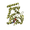
- PDB-3b1x: Crystal structure of an S. thermophilus NFeoB E66A mutant bound t... -

+
Open data


ID or keywords:

Loading...

-
Basic information

Entry
Database: PDB / ID: 3b1x
Title
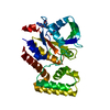
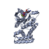
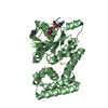
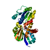
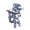
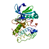
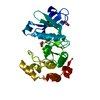
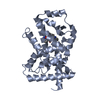
Crystal structure of an S. thermophilus NFeoB E66A mutant bound to GMPPNP
ComponentsFerrous iron uptake transporter protein B
KeywordsMETAL TRANSPORT / G protein / iron transport / GTPase / transmembrane / potassium
Function / homology
Function and homology information


ferrous iron transmembrane transporter activity / GTP binding / metal ion binding / plasma membrane
Similarity search - Function
Helix Hairpins - #1770 / FeoB, cytosolic helical domain / FeoB cytosolic helical domain / Ferrous iron transport protein B, C-terminal / Ferrous iron transport protein B C terminus / Ferrous iron transport protein B / FeoB-type guanine nucleotide-binding (G) domain / Ferrous iron transport protein B / FeoB-type guanine nucleotide-binding (G) domain profile. / Nucleoside transporter/FeoB GTPase, Gate domain ...Helix Hairpins - #1770 / FeoB, cytosolic helical domain / FeoB cytosolic helical domain / Ferrous iron transport protein B, C-terminal / Ferrous iron transport protein B C terminus / Ferrous iron transport protein B / FeoB-type guanine nucleotide-binding (G) domain / Ferrous iron transport protein B / FeoB-type guanine nucleotide-binding (G) domain profile. / Nucleoside transporter/FeoB GTPase, Gate domain / Nucleoside recognition / GTP binding domain / Helix Hairpins / P-loop containing nucleotide triphosphate hydrolases / P-loop containing nucleoside triphosphate hydrolase / Rossmann fold / Orthogonal Bundle / 3-Layer(aba) Sandwich / Mainly Alpha / Alpha Beta
Similarity search - Domain/homology
PHOSPHOAMINOPHOSPHONIC ACID-GUANYLATE ESTER / Ferrous iron transport protein B
Similarity search - Component
Biological speciesStreptococcus thermophilus (bacteria)
MethodX-RAY DIFFRACTION / SYNCHROTRON / MOLECULAR REPLACEMENT / Resolution: 2.61 Å
AuthorsAsh, M.R. / Maher, M.J. / Guss, J.M. / Jormakka, M.
CitationJournal: Acta Crystallogr.,Sect.D / Year: 2011
Title: A suite of Switch I and Switch II mutant structures from the G-protein domain of FeoB
Authors: Ash, M.R. / Maher, M.J. / Guss, J.M. / Jormakka, M.
History
DepositionJul 15, 2011Deposition site: PDBJ / Processing site: PDBJ
Revision 1.0Nov 2, 2011Provider: repository / Type: Initial release
Revision 1.1Apr 18, 2012Group: Database references
Revision 1.2Mar 13, 2024Group: Data collection / Database references / Derived calculations
Category: chem_comp_atom / chem_comp_bond ...chem_comp_atom / chem_comp_bond / database_2 / pdbx_struct_conn_angle / struct_conn / struct_ref_seq_dif / struct_site
Item: _database_2.pdbx_DOI / _database_2.pdbx_database_accession ..._database_2.pdbx_DOI / _database_2.pdbx_database_accession / _pdbx_struct_conn_angle.ptnr1_auth_comp_id / _pdbx_struct_conn_angle.ptnr1_auth_seq_id / _pdbx_struct_conn_angle.ptnr1_label_asym_id / _pdbx_struct_conn_angle.ptnr1_label_atom_id / _pdbx_struct_conn_angle.ptnr1_label_comp_id / _pdbx_struct_conn_angle.ptnr1_label_seq_id / _pdbx_struct_conn_angle.ptnr3_auth_comp_id / _pdbx_struct_conn_angle.ptnr3_auth_seq_id / _pdbx_struct_conn_angle.ptnr3_label_asym_id / _pdbx_struct_conn_angle.ptnr3_label_atom_id / _pdbx_struct_conn_angle.ptnr3_label_comp_id / _pdbx_struct_conn_angle.ptnr3_label_seq_id / _pdbx_struct_conn_angle.value / _struct_conn.pdbx_dist_value / _struct_conn.ptnr1_auth_asym_id / _struct_conn.ptnr1_auth_comp_id / _struct_conn.ptnr1_auth_seq_id / _struct_conn.ptnr1_label_asym_id / _struct_conn.ptnr1_label_atom_id / _struct_conn.ptnr1_label_comp_id / _struct_conn.ptnr1_label_seq_id / _struct_conn.ptnr2_auth_asym_id / _struct_conn.ptnr2_auth_comp_id / _struct_conn.ptnr2_auth_seq_id / _struct_conn.ptnr2_label_asym_id / _struct_conn.ptnr2_label_atom_id / _struct_conn.ptnr2_label_comp_id / _struct_ref_seq_dif.details / _struct_site.pdbx_auth_asym_id / _struct_site.pdbx_auth_comp_id / _struct_site.pdbx_auth_seq_id

-
Structure visualization

Structure viewerMolecule:
MolmilJmol/JSmol

Downloads & links

-
Assembly

Deposited unit
A: Ferrous iron uptake transporter protein B
B: Ferrous iron uptake transporter protein B
hetero molecules


Theoretical massNumber of molelcules
Total (without water)61,3836
Polymers60,2902
Non-polymers1,0934
Water1,02757
1
A: Ferrous iron uptake transporter protein B
hetero molecules


Theoretical massNumber of molelcules
Total (without water)30,6923
Polymers30,1451
Non-polymers5472
Water181
TypeNameSymmetry operationNumber
identity operation1_555x,y,z1
MethodPISA
2
B: Ferrous iron uptake transporter protein B
hetero molecules


Theoretical massNumber of molelcules
Total (without water)30,6923
Polymers30,1451
Non-polymers5472
Water181
TypeNameSymmetry operationNumber
identity operation1_555x,y,z1
MethodPISA
Unit cell
Length a, b, c (Å)48.7, 74.4, 156.1
Angle α, β, γ (deg.)90.000, 90.000, 90.000
Int Tables number19
Space group name H-MP212121

-
Components

#1: Protein Ferrous iron uptake transporter protein B


Mass: 30145.115 Da / Num. of mol.: 2 / Fragment: NFeoB, UNP residues 1-270 / Mutation: E66A
Source method: isolated from a genetically manipulated source
Source: (gene. exp.) Streptococcus thermophilus (bacteria) / Strain: LMG 18311 / Gene: FeoB / Plasmid: pGEX-4T-1 / Production host: Escherichia coli (E. coli) / Strain (production host): BL21(DE3) / References: UniProt: Q5M586
#2: Chemical ChemComp-GNP / PHOSPHOAMINOPHOSPHONIC ACID-GUANYLATE ESTER


Mass: 522.196 Da / Num. of mol.: 2 / Source method: obtained synthetically / Formula: C10H17N6O13P3
Comment: GppNHp, GMPPNP, energy-carrying molecule analogue*YM
#3: Chemical ChemComp-MG / MAGNESIUM ION


Mass: 24.305 Da / Num. of mol.: 2 / Source method: obtained synthetically / Formula: Mg
#4: Water ChemComp-HOH / water


Mass: 18.015 Da / Num. of mol.: 57 / Source method: isolated from a natural source / Formula: H2O

-
Experimental details

-
Experiment

ExperimentMethod: X-RAY DIFFRACTION / Number of used crystals: 1

-
Sample preparation

CrystalDensity Matthews: 2.35 Å3/Da / Density % sol: 47.55 % / Mosaicity: 0.32 °
Crystal growTemperature: 298 K / Method: vapor diffusion, hanging drop / pH: 6
Details: 0.1M SPG buffer, 25%(w/v) PEG 1500, pH 6.0, VAPOR DIFFUSION, HANGING DROP, temperature 298K

-
Data collection

DiffractionMean temperature: 100 K
Diffraction sourceSource: SYNCHROTRON / Site: Australian Synchrotron / Beamline: MX1 / Wavelength: 0.9537 Å
DetectorType: ADSC QUANTUM 210r / Detector: CCD / Date: Feb 3, 2011
RadiationMonochromator: Si(111) / Protocol: SINGLE WAVELENGTH / Monochromatic (M) / Laue (L): M / Scattering type: x-ray
Radiation wavelengthWavelength: 0.9537 Å / Relative weight: 1
ReflectionResolution: 2.6→50 Å / Num. all: 17841 / Num. obs: 17841 / % possible obs: 99.3 % / Redundancy: 5.8 % / Rmerge(I) obs: 0.153 / Χ2: 1.105 / Net I/σ(I): 11.5
Reflection shell

Diffraction-ID: 1

Resolution (Å)Redundancy (%)Rmerge(I) obsMean I/σ(I) obsNum. unique allΧ2% possible all
2.6-2.644.50.5742.17870.88389.4
2.64-2.694.90.5248601.04597.2
2.69-2.745.80.5158680.952100
2.74-2.86.10.4278530.982100
2.8-2.8660.3989281.005100
2.86-2.936.20.3418481.026100
2.93-36.10.3148841.093100
3-3.086.10.2728961.121100
3.08-3.176.10.2378681.157100
3.17-3.2860.29081.107100
3.28-3.396.10.1828771.184100
3.39-3.5360.1638981.212100
3.53-3.6960.1348921.297100
3.69-3.8860.1178871.083100
3.88-4.1360.1028951.169100
4.13-4.455.90.0889191.34100
4.45-4.8960.0849071.057100
4.89-5.65.90.0979151.109100
5.6-7.055.70.1059411.142100
7.05-505.30.06210101.02199.7

-
Processing

Software
NameVersionClassificationNB
SCALEPACKdata scaling
REFMAC5.5.0109refinement
PDB_EXTRACT3.1data extraction
Blu-Icedata collection
HKL-2000data reduction
PHASERphasing
RefinementMethod to determine structure: MOLECULAR REPLACEMENT / Resolution: 2.61→46.5 Å / Cor.coef. Fo:Fc: 0.917 / Cor.coef. Fo:Fc free: 0.865 / Occupancy max: 1 / Occupancy min: 0.5 / SU B: 21.568 / SU ML: 0.235 / Cross valid method: THROUGHOUT / σ(F): 0 / ESU R Free: 0.345 / Stereochemistry target values: MAXIMUM LIKELIHOOD / Details: HYDROGENS HAVE BEEN ADDED IN THE RIDING POSITIONS
RfactorNum. reflection% reflectionSelection details
Rfree0.2591 928 5.2 %RANDOM
Rwork0.214 ---
obs0.2163 17682 98.56 %-
all-17682 --
Solvent computationIon probe radii: 0.8 Å / Shrinkage radii: 0.8 Å / VDW probe radii: 1.4 Å / Solvent model: MASK
Displacement parametersBiso max: 56.2 Å2 / Biso mean: 25.1491 Å2 / Biso min: 5.02 Å2
Baniso -1Baniso -2Baniso -3
1--0.68 Å20 Å20 Å2
2--0.24 Å20 Å2
3---0.43 Å2
Refinement stepCycle: LAST / Resolution: 2.61→46.5 Å
ProteinNucleic acidLigandSolventTotal
Num. atoms4007 0 66 57 4130
Refine LS restraints
Refine-IDTypeDev idealDev ideal targetNumber
X-RAY DIFFRACTIONr_bond_refined_d0.010.0224167
X-RAY DIFFRACTIONr_angle_refined_deg1.4421.9935678
X-RAY DIFFRACTIONr_dihedral_angle_1_deg3.5825522
X-RAY DIFFRACTIONr_dihedral_angle_2_deg33.42125.455187
X-RAY DIFFRACTIONr_dihedral_angle_3_deg12.97915734
X-RAY DIFFRACTIONr_dihedral_angle_4_deg12.3131524
X-RAY DIFFRACTIONr_chiral_restr0.0640.2680
X-RAY DIFFRACTIONr_gen_planes_refined0.0030.0213082
X-RAY DIFFRACTIONr_mcbond_it0.4471.52592
X-RAY DIFFRACTIONr_mcangle_it0.8624216
X-RAY DIFFRACTIONr_scbond_it1.23431575
X-RAY DIFFRACTIONr_scangle_it2.214.51462
LS refinement shellResolution: 2.609→2.677 Å / Total num. of bins used: 20
RfactorNum. reflection% reflection
Rfree0.332 66 -
Rwork0.243 1095 -
all-1161 -
obs-1161 88.97 %
Refinement TLS params.

Method: refined / Refine-ID: X-RAY DIFFRACTION

IDL112)L122)L132)L222)L232)L332)S11 (Å °)S12 (Å °)S13 (Å °)S21 (Å °)S22 (Å °)S23 (Å °)S31 (Å °)S32 (Å °)S33 (Å °)T112)T122)T132)T222)T232)T332)Origin x (Å)Origin y (Å)Origin z (Å)
11.81350.6304-0.77790.5993-0.31920.83830.02670.09620.06960.01080.02250.0283-0.0448-0.0322-0.04910.0540.0189-0.01170.0403-0.00750.0294-3.94517.687-22.331
22.0574-0.8021-0.77090.57830.66710.86090.0040.01460.1417-0.0222-0.0032-0.0051-0.0572-0.0057-0.00080.0707-0.0236-0.00640.05010.01480.0269-21.96614.728-60.943
Refinement TLS group
IDRefine-IDRefine TLS-IDAuth asym-IDAuth seq-ID
1X-RAY DIFFRACTION1A1 - 259
2X-RAY DIFFRACTION2B1 - 257

+
About Yorodumi

-
News

-
Feb 9, 2022. New format data for meta-information of EMDB entries

New format data for meta-information of EMDB entries

  • Version 3 of the EMDB header file is now the official format.
  • The previous official version 1.9 will be removed from the archive.

Related info.:EMDB header

External links:wwPDB to switch to version 3 of the EMDB data model

-
Aug 12, 2020. Covid-19 info

Covid-19 info

URL: https://pdbjlvh1.pdbj.org/emnavi/covid19.php

New page: Covid-19 featured information page in EM Navigator.

Related info.:Covid-19 info / Mar 5, 2020. Novel coronavirus structure data

+
Mar 5, 2020. Novel coronavirus structure data

Novel coronavirus structure data

Related info.:Yorodumi Speices / Aug 12, 2020. Covid-19 info

External links:COVID-19 featured content - PDBj / Molecule of the Month (242):Coronavirus Proteases

+
Jan 31, 2019. EMDB accession codes are about to change! (news from PDBe EMDB page)

EMDB accession codes are about to change! (news from PDBe EMDB page)

  • The allocation of 4 digits for EMDB accession codes will soon come to an end. Whilst these codes will remain in use, new EMDB accession codes will include an additional digit and will expand incrementally as the available range of codes is exhausted. The current 4-digit format prefixed with “EMD-” (i.e. EMD-XXXX) will advance to a 5-digit format (i.e. EMD-XXXXX), and so on. It is currently estimated that the 4-digit codes will be depleted around Spring 2019, at which point the 5-digit format will come into force.
  • The EM Navigator/Yorodumi systems omit the EMD- prefix.

Related info.:Q: What is EMD? / ID/Accession-code notation in Yorodumi/EM Navigator

External links:EMDB Accession Codes are Changing Soon! / Contact to PDBj

+
Jul 12, 2017. Major update of PDB

Major update of PDB

  • wwPDB released updated PDB data conforming to the new PDBx/mmCIF dictionary.
  • This is a major update changing the version number from 4 to 5, and with Remediation, in which all the entries are updated.
  • In this update, many items about electron microscopy experimental information are reorganized (e.g. em_software).
  • Now, EM Navigator and Yorodumi are based on the updated data.

External links:wwPDB Remediation / Enriched Model Files Conforming to OneDep Data Standards Now Available in the PDB FTP Archive

-
Yorodumi

Thousand views of thousand structures

  • Yorodumi is a browser for structure data from EMDB, PDB, SASBDB, etc.
  • This page is also the successor to EM Navigator detail page, and also detail information page/front-end page for Omokage search.
  • The word "yorodu" (or yorozu) is an old Japanese word meaning "ten thousand". "mi" (miru) is to see.

Related info.:EMDB / PDB / SASBDB / Comparison of 3 databanks / Yorodumi Search / Aug 31, 2016. New EM Navigator & Yorodumi / Yorodumi Papers / Jmol/JSmol / Function and homology information / Changes in new EM Navigator and Yorodumi

Read more