 Movie
Movie Controller

[English] 日本語
 Yorodumi
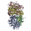
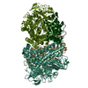
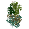
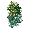
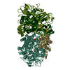
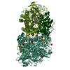
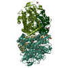
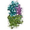
- PDB-3ayz: Membrane-bound respiratory [NiFe] hydrogenase from Hydrogenovibri... -
+ Open data

- Basic information
| Entry | Database: PDB / ID: 3ayz | ||||||
|---|---|---|---|---|---|---|---|
| Title | Membrane-bound respiratory [NiFe] hydrogenase from Hydrogenovibrio marinus in an air-oxidized condition | ||||||
 Components Components | (Membrane-bound hydrogenase ...) x 2 | ||||||
 Keywords Keywords | OXIDOREDUCTASE / MEMBRANE-BOUND NI-FE HYDROGENASE | ||||||
| Function / homology |  Function and homology information Function and homology informationhydrogenase (acceptor) / ferredoxin hydrogenase complex / [Ni-Fe] hydrogenase complex / hydrogenase (acceptor) activity / ferredoxin hydrogenase activity / anaerobic respiration / 3 iron, 4 sulfur cluster binding / nickel cation binding / 4 iron, 4 sulfur cluster binding / electron transfer activity ...hydrogenase (acceptor) / ferredoxin hydrogenase complex / [Ni-Fe] hydrogenase complex / hydrogenase (acceptor) activity / ferredoxin hydrogenase activity / anaerobic respiration / 3 iron, 4 sulfur cluster binding / nickel cation binding / 4 iron, 4 sulfur cluster binding / electron transfer activity / metal ion binding / plasma membrane Similarity search - Function | ||||||
| Biological species |  Hydrogenovibrio marinus (bacteria) Hydrogenovibrio marinus (bacteria) | ||||||
| Method |  X-RAY DIFFRACTION / SYNCHROTRON / MOLECULAR REPLACEMENT / Resolution: 1.22 Å X-RAY DIFFRACTION / SYNCHROTRON / MOLECULAR REPLACEMENT / Resolution: 1.22 Å | ||||||
 Authors Authors | Shomura, Y. / Yoon, K.S. / Nishihara, H. / Higuchi, Y. | ||||||
 Citation Citation | Journal: Nature / Year: 2011 Title: Structural basis for a [4Fe-3S] cluster in the oxygen-tolerant membrane-bound [NiFe]-hydrogenase Authors: Shomura, Y. / Yoon, K.S. / Nishihara, H. / Higuchi, Y. | ||||||
| History |
|
- Structure visualization
| Structure viewer | Molecule: MolmilJmol/JSmol |
|---|
- Downloads & links
-Download
| PDBx/mmCIF format | 3ayz.cif.gz | 769.8 KB | Display | PDBx/mmCIF format |
|---|---|---|---|---|
| PDB format | pdb3ayz.ent.gz | 621.4 KB | Display | PDB format |
| PDBx/mmJSON format | 3ayz.json.gz | Tree view | PDBx/mmJSON format | |
| Others |  Other downloads Other downloads |
-Validation report
| Arichive directory | https://data.pdbj.org/pub/pdb/validation_reports/ay/3ayzftp://data.pdbj.org/pub/pdb/validation_reports/ay/3ayz | HTTPS FTP |
|---|
-Related structure data
| Related structure data |  3ayxSC  5y34C S: Starting model for refinement C: citing same article ( |
|---|---|
| Similar structure data |


-Links
 PDBj
PDBj


- Assembly
| Deposited unit | 
| ||||||||
|---|---|---|---|---|---|---|---|---|---|
| 1 |
| ||||||||
| Unit cell |
|
-Components
-Membrane-bound hydrogenase ... , 2 types, 4 molecules ACBD
| #1: Protein | Mass: 66574.398 Da / Num. of mol.: 2 / Source method: isolated from a natural source / Source: (natural) Hydrogenovibrio marinus (bacteria) / Strain: MH-110 / References: UniProt: F2Z6J6, EC: 1.12.5.1#2: Protein | Mass: 31306.662 Da / Num. of mol.: 2 / Fragment: UNP residues 41-323 / Source method: isolated from a natural source / Source: (natural) Hydrogenovibrio marinus (bacteria) / Strain: MH-110 / References: UniProt: F2Z6J5, EC: 1.12.5.1 |
|---|
-Non-polymers , 12 types, 1665 molecules 


| #3: Chemical |  Mass: 55.845 Da / Num. of mol.: 2 / Source method: obtained synthetically / Formula: Fe Mass: 55.845 Da / Num. of mol.: 2 / Source method: obtained synthetically / Formula: Fe#4: Chemical |  Mass: 58.693 Da / Num. of mol.: 2 / Source method: obtained synthetically / Formula: Ni Mass: 58.693 Da / Num. of mol.: 2 / Source method: obtained synthetically / Formula: Ni#5: Chemical |  Mass: 28.010 Da / Num. of mol.: 2 / Source method: obtained synthetically / Formula: CO Mass: 28.010 Da / Num. of mol.: 2 / Source method: obtained synthetically / Formula: CO#6: Chemical | ChemComp-CYN /  Mass: 26.017 Da / Num. of mol.: 4 / Source method: obtained synthetically / Formula: CN Mass: 26.017 Da / Num. of mol.: 4 / Source method: obtained synthetically / Formula: CN#7: Chemical | ChemComp-O /  Mass: 15.999 Da / Num. of mol.: 10 / Source method: obtained synthetically / Formula: O Mass: 15.999 Da / Num. of mol.: 10 / Source method: obtained synthetically / Formula: O#8: Chemical | ChemComp-GOL /  Mass: 92.094 Da / Num. of mol.: 4 / Source method: obtained synthetically / Formula: C3H8O3 Mass: 92.094 Da / Num. of mol.: 4 / Source method: obtained synthetically / Formula: C3H8O3#9: Chemical |  Mass: 24.305 Da / Num. of mol.: 2 / Source method: obtained synthetically / Formula: Mg Mass: 24.305 Da / Num. of mol.: 2 / Source method: obtained synthetically / Formula: Mg#10: Chemical |  Mass: 319.575 Da / Num. of mol.: 2 / Source method: obtained synthetically / Formula: Fe4S3 Mass: 319.575 Da / Num. of mol.: 2 / Source method: obtained synthetically / Formula: Fe4S3#11: Chemical |  Mass: 319.575 Da / Num. of mol.: 2 / Source method: obtained synthetically / Formula: Fe4S3 Mass: 319.575 Da / Num. of mol.: 2 / Source method: obtained synthetically / Formula: Fe4S3#12: Chemical |  Mass: 295.795 Da / Num. of mol.: 2 / Source method: obtained synthetically / Formula: Fe3S4 Mass: 295.795 Da / Num. of mol.: 2 / Source method: obtained synthetically / Formula: Fe3S4#13: Chemical |  Mass: 351.640 Da / Num. of mol.: 2 / Source method: obtained synthetically / Formula: Fe4S4 Mass: 351.640 Da / Num. of mol.: 2 / Source method: obtained synthetically / Formula: Fe4S4#14: Water | ChemComp-HOH / | Mass: 18.015 Da / Num. of mol.: 1631 / Source method: isolated from a natural source / Formula: H2O |
|---|
-Experimental details
-Experiment
| Experiment | Method: X-RAY DIFFRACTION / Number of used crystals: 1 |
|---|
- Sample preparation
| Crystal | Density Matthews: 2.56 Å3/Da / Density % sol: 51.94 % Description: THE STRUCTURE FACTOR FILE CONTAINS FRIEDEL PAIRS |
|---|---|
| Crystal grow | Temperature: 293 K / Method: vapor diffusion, sitting drop / pH: 6.5 Details: 0.1M PIPES, 15% PEG 3350, 300mM lithium sulfate, 5mM DTT, pH 6.5, VAPOR DIFFUSION, SITTING DROP, temperature 293K |
-Data collection
| Diffraction | Mean temperature: 90 K |
|---|---|
| Diffraction source | Source: SYNCHROTRON / Site: SPring-8  / Beamline: BL44XU / Wavelength: 0.9 Å / Beamline: BL44XU / Wavelength: 0.9 Å |
| Detector | Type: RAYONIX MX225HE / Detector: CCD / Date: Oct 21, 2010 |
| Radiation | Monochromator: SI(111) / Protocol: SINGLE WAVELENGTH / Monochromatic (M) / Laue (L): M / Scattering type: x-ray |
| Radiation wavelength | Wavelength: 0.9 Å / Relative weight: 1 |
| Reflection | Resolution: 1.22→20 Å / Num. all: 582417 / Num. obs: 578159 / % possible obs: 99.6 % / Redundancy: 4.1 % / Biso Wilson estimate: 10.3 Å2 / Rmerge(I) obs: 0.084 / Net I/σ(I): 12.5 |
| Reflection shell | Resolution: 1.22→1.24 Å / Redundancy: 3.2 % / Rmerge(I) obs: 0.385 / Mean I/σ(I) obs: 3.3 / % possible all: 99.4 |
- Processing
| Software |
| |||||||||||||||||||||||||||||||||
|---|---|---|---|---|---|---|---|---|---|---|---|---|---|---|---|---|---|---|---|---|---|---|---|---|---|---|---|---|---|---|---|---|---|---|
| Refinement | Method to determine structure: MOLECULAR REPLACEMENT Starting model: PDB ENTRY 3AYX Resolution: 1.22→20 Å / Num. parameters: 140969 / Num. restraintsaints: 174515 / Cross valid method: FREE R / σ(F): 0 / Stereochemistry target values: Engh & Huber Details: 1. The SF file contains friedel pairs. Friedel pairs were independently treated during refinement. Statistics for data collection were calculated from data of which the friedel pairs were ...Details: 1. The SF file contains friedel pairs. Friedel pairs were independently treated during refinement. Statistics for data collection were calculated from data of which the friedel pairs were merged. 2. ANISOTROPIC SCALING APPLIED BY THE METHOD OF PARKIN, MOEZZI & HOPE, J.APPL.CRYST.28(1995)53-56 ANISOTROPIC REFINEMENT REDUCED FREE R (NO CUTOFF) BY ?
| |||||||||||||||||||||||||||||||||
| Refine analyze | Num. disordered residues: 52 / Occupancy sum hydrogen: 13111 / Occupancy sum non hydrogen: 15255.7 | |||||||||||||||||||||||||||||||||
| Refinement step | Cycle: LAST / Resolution: 1.22→20 Å
| |||||||||||||||||||||||||||||||||
| Refine LS restraints |
|
