 Movie
Movie Controller

[English] 日本語
 Yorodumi
- PDB-3avj: Crystal structures of novel allosteric peptide inhibitors of HIV ... -
+ Open data

- Basic information
| Entry | Database: PDB / ID: 3avj | ||||||
|---|---|---|---|---|---|---|---|
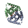
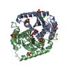
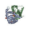
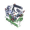
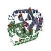
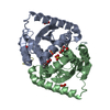
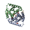
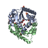
| Title | Crystal structures of novel allosteric peptide inhibitors of HIV integrase in the LEDGF binding site | ||||||
 Components Components |
| ||||||
 Keywords Keywords | RECOMBINATION/INHIBITOR / protein-protein interactions / HIV / RECOMBINATION-INHIBITOR complex | ||||||
| Function / homology |  Function and homology information Function and homology informationHIV-1 retropepsin / symbiont-mediated activation of host apoptosis / retroviral ribonuclease H / exoribonuclease H / exoribonuclease H activity / DNA integration / viral genome integration into host DNA / establishment of integrated proviral latency / RNA-directed DNA polymerase / RNA stem-loop binding ...HIV-1 retropepsin / symbiont-mediated activation of host apoptosis / retroviral ribonuclease H / exoribonuclease H / exoribonuclease H activity / DNA integration / viral genome integration into host DNA / establishment of integrated proviral latency / RNA-directed DNA polymerase / RNA stem-loop binding / viral penetration into host nucleus / host multivesicular body / RNA-directed DNA polymerase activity / RNA-DNA hybrid ribonuclease activity / Transferases; Transferring phosphorus-containing groups; Nucleotidyltransferases / host cell / viral nucleocapsid / DNA recombination / DNA-directed DNA polymerase / aspartic-type endopeptidase activity / Hydrolases; Acting on ester bonds / DNA-directed DNA polymerase activity / symbiont-mediated suppression of host gene expression / viral translational frameshifting / symbiont entry into host cell / lipid binding / host cell nucleus / host cell plasma membrane / virion membrane / structural molecule activity / proteolysis / DNA binding / zinc ion binding Similarity search - Function | ||||||
| Biological species |   Human immunodeficiency virus type 1 Human immunodeficiency virus type 1 Homo sapiens (human) Homo sapiens (human) | ||||||
| Method |  X-RAY DIFFRACTION / SYNCHROTRON / MOLECULAR REPLACEMENT / molecular replacement / Resolution: 1.7 Å X-RAY DIFFRACTION / SYNCHROTRON / MOLECULAR REPLACEMENT / molecular replacement / Resolution: 1.7 Å | ||||||
 Authors Authors | Peat, T.S. / Deadman, J.J. / Newman, J. / Rhodes, D.I. | ||||||
 Citation Citation | Journal: Chembiochem / Year: 2011 Title: Crystal structures of novel allosteric peptide inhibitors of HIV integrase identify new interactions at the LEDGF binding site. Authors: Rhodes, D.I. / Peat, T.S. / Vandegraaff, N. / Jeevarajah, D. / Newman, J. / Martyn, J. / Coates, J.A. / Ede, N.J. / Rea, P. / Deadman, J.J. | ||||||
| History |
|
- Structure visualization
| Structure viewer | Molecule: MolmilJmol/JSmol |
|---|
- Downloads & links
-Download
| PDBx/mmCIF format | 3avj.cif.gz | 86.6 KB | Display | PDBx/mmCIF format |
|---|---|---|---|---|
| PDB format | pdb3avj.ent.gz | 65.6 KB | Display | PDB format |
| PDBx/mmJSON format | 3avj.json.gz | Tree view | PDBx/mmJSON format | |
| Others |  Other downloads Other downloads |
-Validation report
| Arichive directory | https://data.pdbj.org/pub/pdb/validation_reports/av/3avjftp://data.pdbj.org/pub/pdb/validation_reports/av/3avj | HTTPS FTP |
|---|
-Related structure data
| Related structure data | 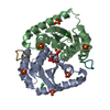 3av9C  3avaC  3avbC  3avcC  3avfC  3avgC  3avhC  3aviC 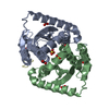 3avkC 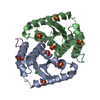 3avlC 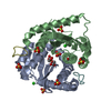 3avmC 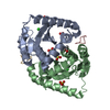 3avnC C: citing same article ( |
|---|---|
| Similar structure data |


-Links
 PDBj
PDBj


- Assembly
| Deposited unit | 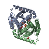
| ||||||||
|---|---|---|---|---|---|---|---|---|---|
| 1 |
| ||||||||
| Unit cell |
|
-Components
-Protein / Protein/peptide , 2 types, 4 molecules ABDF
| #1: Protein | Mass: 20044.672 Da / Num. of mol.: 2 / Fragment: CCD domain (UNP RESIDUES 1197-1359) / Mutation: C56S, F139D, F185H Source method: isolated from a genetically manipulated source Source: (gene. exp.) Human immunodeficiency virus type 1 / Strain: NEW YORK-5 ISOLATE / Gene: gag-pol / Plasmid: pET28 / Production host:  #2: Protein/peptide |   Type: Cyclic peptide / Class: Enzyme inhibitor / Mass: 920.063 Da / Num. of mol.: 2 / Source method: obtained synthetically / Details: chemical synthsis / Source: (synth.) Homo sapiens (human) / References: ALKIDNMD peptide Type: Cyclic peptide / Class: Enzyme inhibitor / Mass: 920.063 Da / Num. of mol.: 2 / Source method: obtained synthetically / Details: chemical synthsis / Source: (synth.) Homo sapiens (human) / References: ALKIDNMD peptide |
|---|
-Non-polymers , 4 types, 197 molecules 


| #3: Chemical | ChemComp-SO4 /  Mass: 96.063 Da / Num. of mol.: 8 / Source method: obtained synthetically / Formula: SO4 Mass: 96.063 Da / Num. of mol.: 8 / Source method: obtained synthetically / Formula: SO4#4: Chemical |  Mass: 35.453 Da / Num. of mol.: 2 / Source method: obtained synthetically / Formula: Cl Mass: 35.453 Da / Num. of mol.: 2 / Source method: obtained synthetically / Formula: Cl#5: Chemical |  Mass: 60.052 Da / Num. of mol.: 3 / Source method: obtained synthetically / Formula: C2H4O2 Mass: 60.052 Da / Num. of mol.: 3 / Source method: obtained synthetically / Formula: C2H4O2#6: Water | ChemComp-HOH / | Mass: 18.015 Da / Num. of mol.: 184 / Source method: isolated from a natural source / Formula: H2O |
|---|
-Details
| Has protein modification | Y |
|---|
-Experimental details
-Experiment
| Experiment | Method: X-RAY DIFFRACTION / Number of used crystals: 1 |
|---|
- Sample preparation
| Crystal | Density Matthews: 2.33 Å3/Da / Density % sol: 47.14 % |
|---|---|
| Crystal grow | Temperature: 293 K / Method: vapor diffusion, sitting drop Details: 40mM Tris pH 8.0, 250mM NaCl, 30mM MgCl2, 5mM DTT, 1.6-2.0M ammonium sulfate, 100mM sodium acetate buffer pH 5.0-5.5, vapor diffusion, sitting drop, temperature 293K PH range: 5.0-5.5 |
-Data collection
| Diffraction | Mean temperature: 100 K |
|---|---|
| Diffraction source | Source: SYNCHROTRON / Site: Australian Synchrotron  / Beamline: MX2 / Wavelength: 0.96 Å / Beamline: MX2 / Wavelength: 0.96 Å |
| Detector | Type: ADSC QUANTUM 315 / Detector: CCD / Date: Dec 14, 2009 |
| Radiation | Protocol: SINGLE WAVELENGTH / Monochromatic (M) / Laue (L): M / Scattering type: x-ray |
| Radiation wavelength | Wavelength: 0.96 Å / Relative weight: 1 |
| Reflection | Resolution: 1.7→67 Å / Num. all: 41624 / Num. obs: 41624 |
-Phasing
| Phasing | Method: molecular replacement |
|---|
- Processing
| Software |
| |||||||||||||||||||||||||||||||||||||||||||||
|---|---|---|---|---|---|---|---|---|---|---|---|---|---|---|---|---|---|---|---|---|---|---|---|---|---|---|---|---|---|---|---|---|---|---|---|---|---|---|---|---|---|---|---|---|---|---|
| Refinement | Method to determine structure: MOLECULAR REPLACEMENT / Resolution: 1.7→61.5 Å / Cor.coef. Fo:Fc: 0.968 / Cor.coef. Fo:Fc free: 0.95 / Occupancy max: 1 / Occupancy min: 0.3 / SU B: 1.536 / SU ML: 0.052 / Cross valid method: THROUGHOUT / σ(F): 0 / ESU R Free: 0.093 / Stereochemistry target values: MAXIMUM LIKELIHOOD Details: HYDROGENS HAVE BEEN USED IF PRESENT IN THE INPUT U VALUES: REFINED INDIVIDUALLY
| |||||||||||||||||||||||||||||||||||||||||||||
| Solvent computation | Ion probe radii: 0.8 Å / Shrinkage radii: 0.8 Å / VDW probe radii: 1.2 Å / Solvent model: MASK | |||||||||||||||||||||||||||||||||||||||||||||
| Displacement parameters | Biso max: 76.19 Å2 / Biso mean: 22.5965 Å2 / Biso min: 7.37 Å2
| |||||||||||||||||||||||||||||||||||||||||||||
| Refinement step | Cycle: LAST / Resolution: 1.7→61.5 Å
| |||||||||||||||||||||||||||||||||||||||||||||
| Refine LS restraints |
| |||||||||||||||||||||||||||||||||||||||||||||
| LS refinement shell | Resolution: 1.7→1.744 Å / Total num. of bins used: 20
|
