 Movie
Movie Controller

[English] 日本語
 Yorodumi
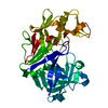
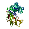
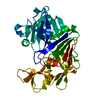
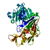
- PDB-3app: STRUCTURE AND REFINEMENT OF PENICILLOPEPSIN AT 1.8 ANGSTROMS RESO... -
+ Open data

- Basic information
| Entry | Database: PDB / ID: 3app | |||||||||
|---|---|---|---|---|---|---|---|---|---|---|
| Title | STRUCTURE AND REFINEMENT OF PENICILLOPEPSIN AT 1.8 ANGSTROMS RESOLUTION | |||||||||
 Components Components | PENICILLOPEPSIN | |||||||||
 Keywords Keywords | HYDROLASE (ACID PROTEINASE) | |||||||||
| Function / homology |  Function and homology information Function and homology informationpenicillopepsin / aspartic-type endopeptidase activity / proteolysis / extracellular region Similarity search - Function | |||||||||
| Biological species |  Penicillium janthinellum (fungus) Penicillium janthinellum (fungus) | |||||||||
| Method |  X-RAY DIFFRACTION / Resolution: 1.8 Å X-RAY DIFFRACTION / Resolution: 1.8 Å | |||||||||
 Authors Authors | Sielecki, A.R. / James, M.N.G. | |||||||||
 Citation Citation | Journal: J.Mol.Biol. / Year: 1983 Title: Structure and refinement of penicillopepsin at 1.8 A resolution. Authors: James, M.N. / Sielecki, A.R. #1: Journal: Biological Macromolecules and Assemblies / Year: 1987Title: Aspartic Proteinases and Their Catalytic Pathway Authors: James, M.N.G. / Sielecki, A.R. #2: Journal: Biochemistry / Year: 1985Title: Stereochemical Analysis of Peptide Bond Hydrolysis Catalyzed by the Aspartic Proteinase Penicillopepsin Authors: James, M.N.G. / Sielecki, A.R. #3: Journal: Proc.Natl.Acad.Sci.USA / Year: 1982Title: Conformational Flexibility in the Active Sites of Aspartyl Proteinases Revealed by a Pepstatin Fragment Binding to Penicillopepsin Authors: James, M.N.G. / Sielecki, A. / Salituro, F. / Rich, D.H. / Hofmann, T. #4: Journal: STRUCTURAL STUDIES ON MOLECULES OF BIOLOGICA INTERESTLYear: 1981 Title: The Tertiary Structure of Penicillopepsin. Towards a Catalytic Mechanism for Acid Proteases Authors: James, M.N.G. / Hsu, I-N. / Hofmann, T. / Sielecki, A.R. #5: Journal: Can.J.Biochem. / Year: 1980Title: An X-Ray Crystallographic Approach to Enzyme Structure and Function Authors: James, M.N.G. #6: Journal: Nature / Year: 1978Title: Structural Evidence for Gene Duplication in the Evolution of the Acid Proteases Authors: Tang, J. / James, M.N.G. / Hsu, I.N. / Jenkins, J.A. / Blundell, T.L. #7: Journal: Nature / Year: 1977Title: Mechanism of Acid Protease Catalysis Based on the Crystal Structure of Penicillopepsin Authors: James, M.N.G. / Hsu, I.-N. / Delbaere, L.T.J. #8: Journal: Nature / Year: 1977Title: Penicillopepsin from Penicillium Janthinellum Crystal Structure at 2.8 Angstroms and Sequence Homology with Porcine Pepsin Authors: Hsu, I.-N. / Delbaere, L.T.J. / James, M.N.G. / Hofmann, T. #9: Journal: Adv.Exp.Med.Biol. / Year: 1977Title: Penicillopepsin. 2.8 Angstroms Structure, Active Site Conformation and Mechanistic Implications Authors: Hsu, I-N. / Delbaere, L.T.J. / James, M.N.G. / Hofmann, T. #10: Journal: Biochem.Biophys.Res.Commun. / Year: 1976Title: The Crystal Structure of Penicillopepsin at 6 Angstroms Resolution Authors: Hsu, I-N. / Hofmann, T. / Nyburg, S.C. / James, M.N.G. | |||||||||
| History |
| |||||||||
| Remark 650 | HELIX THE HYDROGEN-BONDING PATTERN AND THE VALUES OF THE PHI, PSI ANGLES WERE USED TO IDENTIFY THE ...HELIX THE HYDROGEN-BONDING PATTERN AND THE VALUES OF THE PHI, PSI ANGLES WERE USED TO IDENTIFY THE SEVERAL SHORT SEGMENTS OF ALPHA-HELICAL CONFORMATION. THERE ARE SIX ALPHA-HELICES, RANGING IN LENGTH FROM 1 TO 2.5 TURNS. FOUR OF THEM HAVE ASSOCIATED 3/10 HELICES INITIATING AND/OR TERMINATING THEM. THE TWO SINGLE TURNS OF ALPHA-HELIX, PRO 59 - GLY 63, ASP 239 - GLY 243, ARE IRREGULAR. THEIR ASSIGNMENT AS ALPHA-HELICAL WAS MADE ON THE BASIS OF GOOD 5-1 HYDROGEN BONDING. THE SPECIFICS ON 3/10 HELICES ARE AS FOLLOWS H1. ASN 58 - ALA 61 3/10 HELIX TYPE III H2. SER 109 - PHE 112 3/10 HELIX TYPE III PHE 112 - ASP 115 3/10 HELIX TYPE III H3. VAL 144 - SER 147 3/10 HELIX TYPE III LYS 145 - LEU 148 3/10 HELIX TYPE I H4. ASP 222 - VAL 225 3/10 HELIX TYPE III TYR 229 - GLN 232 3/10 HELIX TYPE III TYR 230 - VAL 233 3/10 HELIX TYPE I H6. GLY 299 - PHE 302 3/10 HELIX TYPE III PHE 302 - SER 305 3/10 HELIX TYPE III LEU 303 - GLN 306 3/10 HELIX TYPE I |
- Structure visualization
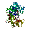
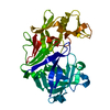
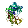
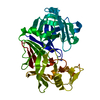
| Structure viewer | Molecule: MolmilJmol/JSmol |
|---|
- Downloads & links
-Download
| PDBx/mmCIF format | 3app.cif.gz | 81.7 KB | Display | PDBx/mmCIF format |
|---|---|---|---|---|
| PDB format | pdb3app.ent.gz | 57.8 KB | Display | PDB format |
| PDBx/mmJSON format | 3app.json.gz | Tree view | PDBx/mmJSON format | |
| Others |  Other downloads Other downloads |
-Validation report
| Arichive directory | https://data.pdbj.org/pub/pdb/validation_reports/ap/3appftp://data.pdbj.org/pub/pdb/validation_reports/ap/3app | HTTPS FTP |
|---|
-Related structure data
| Similar structure data |
|---|


-Links
 PDBj
PDBj
- Assembly
| Deposited unit | 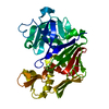
| ||||||||
|---|---|---|---|---|---|---|---|---|---|
| 1 |
| ||||||||
| Unit cell |
| ||||||||
| Atom site foot note | 1: THE REGION FROM SER 277 TO SER 281 IS POORLY ORDERED IN THE ELECTRON DENSITY MAP AT CYCLE 86 (SEE FIGURE 6C IN THE PAPER CITED AS REFERENCE 1 ABOVE). 2: RESIDUES 134 AND 315 ARE CIS-PROLINES. |
-Components
| #1: Protein | Mass: 33468.809 Da / Num. of mol.: 1 Source method: isolated from a genetically manipulated source Source: (gene. exp.) Penicillium janthinellum (fungus) / References: UniProt: P00798, penicillopepsin |
|---|---|
| #2: Water | ChemComp-HOH /  Mass: 18.015 Da / Num. of mol.: 318 / Source method: isolated from a natural source / Formula: H2O Mass: 18.015 Da / Num. of mol.: 318 / Source method: isolated from a natural source / Formula: H2O |
| Has protein modification | Y |
-Experimental details
-Experiment
| Experiment | Method: X-RAY DIFFRACTION |
|---|
- Sample preparation
| Crystal | Density Matthews: 2.01 Å3/Da / Density % sol: 38.67 % | ||||||||||||||||||||
|---|---|---|---|---|---|---|---|---|---|---|---|---|---|---|---|---|---|---|---|---|---|
| Crystal grow | *PLUS pH: 4.4 / Method: unknown | ||||||||||||||||||||
| Components of the solutions | *PLUS
|
- Processing
| Software | Name: PROLSQ / Classification: refinement | ||||||||||||
|---|---|---|---|---|---|---|---|---|---|---|---|---|---|
| Refinement | Resolution: 1.8→8 Å / σ(I): 3 Details: THE REGION FROM SER 277 TO SER 281 IS POORLY ORDERED IN THE ELECTRON DENSITY
| ||||||||||||
| Refinement step | Cycle: LAST / Resolution: 1.8→8 Å
| ||||||||||||
| Software | *PLUS Name: PROLSQ / Classification: refinement | ||||||||||||
| Refinement | *PLUS Rfactor obs: 0.126 | ||||||||||||
| Solvent computation | *PLUS | ||||||||||||
| Displacement parameters | *PLUS |
