 Movie
Movie Controller

[English] 日本語
 Yorodumi
- PDB-3aeu: Structure of the light-independent protochlorophyllide reductase ... -
+ Open data

- Basic information
| Entry | Database: PDB / ID: 3aeu | ||||||
|---|---|---|---|---|---|---|---|
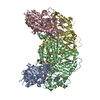
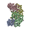
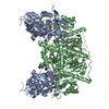
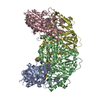
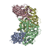
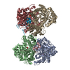
| Title | Structure of the light-independent protochlorophyllide reductase catalyzing a key reduction for greening in the dark | ||||||
 Components Components |
| ||||||
 Keywords Keywords | OXIDOREDUCTASE / Iron/sulfur cluster / Bacteriochlorophyll biosynthesis / Chlorophyll biosynthesis / Photosynthesis | ||||||
| Function / homology |  Function and homology information Function and homology informationferredoxin:protochlorophyllide reductase (ATP-dependent) / photosynthesis, dark reaction / light-independent bacteriochlorophyll biosynthetic process / oxidoreductase activity, acting on iron-sulfur proteins as donors / oxidoreductase activity, acting on the CH-CH group of donors, iron-sulfur protein as acceptor / 4 iron, 4 sulfur cluster binding / ATP binding / metal ion binding Similarity search - Function | ||||||
| Biological species |  Rhodobacter capsulatus (bacteria) Rhodobacter capsulatus (bacteria) | ||||||
| Method |  X-RAY DIFFRACTION / SYNCHROTRON / MOLECULAR REPLACEMENT / Resolution: 2.9 Å X-RAY DIFFRACTION / SYNCHROTRON / MOLECULAR REPLACEMENT / Resolution: 2.9 Å | ||||||
 Authors Authors | Muraki, N. / Nomata, J. / Shiba, T. / Fujita, Y. / Kurisu, G. | ||||||
 Citation Citation | Journal: Nature / Year: 2010 Title: X-ray crystal structure of the light-independent protochlorophyllide reductase Authors: Muraki, N. / Nomata, J. / Ebata, K. / Mizoguchi, T. / Shiba, T. / Tamiaki, H. / Kurisu, G. / Fujita, Y. | ||||||
| History |
|
- Structure visualization
| Structure viewer | Molecule: MolmilJmol/JSmol |
|---|
- Downloads & links
-Download
| PDBx/mmCIF format | 3aeu.cif.gz | 325.5 KB | Display | PDBx/mmCIF format |
|---|---|---|---|---|
| PDB format | pdb3aeu.ent.gz | 258.7 KB | Display | PDB format |
| PDBx/mmJSON format | 3aeu.json.gz | Tree view | PDBx/mmJSON format | |
| Others |  Other downloads Other downloads |
-Validation report
| Arichive directory | https://data.pdbj.org/pub/pdb/validation_reports/ae/3aeuftp://data.pdbj.org/pub/pdb/validation_reports/ae/3aeu | HTTPS FTP |
|---|
-Related structure data
| Related structure data |  3aekC  3aeqC  3aerC  3aesC  3aetC  2zmp C: citing same article ( S: Starting model for refinement |
|---|---|
| Similar structure data |


-Links
 PDBj
PDBj
- Assembly
| Deposited unit | 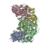
| ||||||||||||||||||||||||||||||||||||||||||||||||||||||||||||||||||||
|---|---|---|---|---|---|---|---|---|---|---|---|---|---|---|---|---|---|---|---|---|---|---|---|---|---|---|---|---|---|---|---|---|---|---|---|---|---|---|---|---|---|---|---|---|---|---|---|---|---|---|---|---|---|---|---|---|---|---|---|---|---|---|---|---|---|---|---|---|---|
| 1 |
| ||||||||||||||||||||||||||||||||||||||||||||||||||||||||||||||||||||
| Unit cell |
| ||||||||||||||||||||||||||||||||||||||||||||||||||||||||||||||||||||
| Noncrystallographic symmetry (NCS) | NCS domain:
NCS domain segments: Component-ID: 1 / Refine code: 3
NCS ensembles :
|
-Components
| #1: Protein | Mass: 47212.086 Da / Num. of mol.: 2 Source method: isolated from a genetically manipulated source Source: (gene. exp.) Rhodobacter capsulatus (bacteria) / Gene: bchN / Plasmid: PASK-IBA5PLUS / Production host: References: UniProt: P26164, Oxidoreductases; Acting on iron-sulfur proteins as donors #2: Protein | Mass: 57217.520 Da / Num. of mol.: 2 / Mutation: D36A Source method: isolated from a genetically manipulated source Source: (gene. exp.) Rhodobacter capsulatus (bacteria) / Gene: bchB, bchK / Plasmid: PASK-IBA5PLUS / Production host: References: UniProt: P26163, Oxidoreductases; Acting on iron-sulfur proteins as donors #3: Chemical |   Mass: 351.640 Da / Num. of mol.: 2 / Source method: obtained synthetically / Formula: Fe4S4 Mass: 351.640 Da / Num. of mol.: 2 / Source method: obtained synthetically / Formula: Fe4S4#4: Water | ChemComp-HOH / |  Mass: 18.015 Da / Num. of mol.: 15 / Source method: isolated from a natural source / Formula: H2O Mass: 18.015 Da / Num. of mol.: 15 / Source method: isolated from a natural source / Formula: H2O |
|---|
-Experimental details
-Experiment
| Experiment | Method: X-RAY DIFFRACTION / Number of used crystals: 1 |
|---|
- Sample preparation
| Crystal | Density Matthews: 2.71 Å3/Da / Density % sol: 54.68 % |
|---|---|
| Crystal grow | Temperature: 277 K / Method: vapor diffusion, hanging drop / pH: 8 Details: 20% PEG3350, 0.2M SODIUM CLORIDE, 0.1M MOPS-NAOH(PH7.0), pH 8.0, VAPOR DIFFUSION, HANGING DROP, temperature 277K |
-Data collection
| Diffraction | Mean temperature: 100 K |
|---|---|
| Diffraction source | Source: SYNCHROTRON / Site: Photon Factory  / Beamline: AR-NW12A / Wavelength: 1 / Beamline: AR-NW12A / Wavelength: 1 |
| Detector | Type: ADSC QUANTUM 210 / Detector: CCD / Date: Feb 6, 2008 |
| Radiation | Protocol: SINGLE WAVELENGTH / Monochromatic (M) / Laue (L): M / Scattering type: x-ray |
| Radiation wavelength | Wavelength: 1 Å / Relative weight: 1 |
| Reflection | Resolution: 2.9→50 Å / Num. obs: 48730 / % possible obs: 97.9 % / Observed criterion σ(I): -2 / Biso Wilson estimate: 63.03 Å2 / Rmerge(I) obs: 0.141 |
- Processing
| Software |
| ||||||||||||||||||||||||||||||||||||||||||||||||||||||||||||||||||||||||||||||||||||||||||||||||||||||||||||||||||||||||||||||||||||||||||||||||||||||||||||||||||||||||||
|---|---|---|---|---|---|---|---|---|---|---|---|---|---|---|---|---|---|---|---|---|---|---|---|---|---|---|---|---|---|---|---|---|---|---|---|---|---|---|---|---|---|---|---|---|---|---|---|---|---|---|---|---|---|---|---|---|---|---|---|---|---|---|---|---|---|---|---|---|---|---|---|---|---|---|---|---|---|---|---|---|---|---|---|---|---|---|---|---|---|---|---|---|---|---|---|---|---|---|---|---|---|---|---|---|---|---|---|---|---|---|---|---|---|---|---|---|---|---|---|---|---|---|---|---|---|---|---|---|---|---|---|---|---|---|---|---|---|---|---|---|---|---|---|---|---|---|---|---|---|---|---|---|---|---|---|---|---|---|---|---|---|---|---|---|---|---|---|---|---|---|---|
| Refinement | Method to determine structure: MOLECULAR REPLACEMENT Starting model: 2ZMP 2zmp Resolution: 2.9→39.9 Å / Cor.coef. Fo:Fc: 0.921 / Cor.coef. Fo:Fc free: 0.876 / SU B: 32.579 / SU ML: 0.285 / TLS residual ADP flag: LIKELY RESIDUAL / Cross valid method: THROUGHOUT / ESU R Free: 0.402 / Stereochemistry target values: MAXIMUM LIKELIHOOD / Details: HYDROGENS HAVE BEEN ADDED IN THE RIDING POSITIONS
| ||||||||||||||||||||||||||||||||||||||||||||||||||||||||||||||||||||||||||||||||||||||||||||||||||||||||||||||||||||||||||||||||||||||||||||||||||||||||||||||||||||||||||
| Solvent computation | Ion probe radii: 0.8 Å / Shrinkage radii: 0.8 Å / VDW probe radii: 1.2 Å / Solvent model: MASK | ||||||||||||||||||||||||||||||||||||||||||||||||||||||||||||||||||||||||||||||||||||||||||||||||||||||||||||||||||||||||||||||||||||||||||||||||||||||||||||||||||||||||||
| Displacement parameters | Biso mean: 26 Å2
| ||||||||||||||||||||||||||||||||||||||||||||||||||||||||||||||||||||||||||||||||||||||||||||||||||||||||||||||||||||||||||||||||||||||||||||||||||||||||||||||||||||||||||
| Refinement step | Cycle: LAST / Resolution: 2.9→39.9 Å
| ||||||||||||||||||||||||||||||||||||||||||||||||||||||||||||||||||||||||||||||||||||||||||||||||||||||||||||||||||||||||||||||||||||||||||||||||||||||||||||||||||||||||||
| Refine LS restraints |
| ||||||||||||||||||||||||||||||||||||||||||||||||||||||||||||||||||||||||||||||||||||||||||||||||||||||||||||||||||||||||||||||||||||||||||||||||||||||||||||||||||||||||||
| Refine LS restraints NCS | Dom-ID: 1 / Refine-ID: X-RAY DIFFRACTION
| ||||||||||||||||||||||||||||||||||||||||||||||||||||||||||||||||||||||||||||||||||||||||||||||||||||||||||||||||||||||||||||||||||||||||||||||||||||||||||||||||||||||||||
| LS refinement shell | Resolution: 2.9→2.98 Å / Total num. of bins used: 20
| ||||||||||||||||||||||||||||||||||||||||||||||||||||||||||||||||||||||||||||||||||||||||||||||||||||||||||||||||||||||||||||||||||||||||||||||||||||||||||||||||||||||||||
| Refinement TLS params. | Method: refined / Refine-ID: X-RAY DIFFRACTION
| ||||||||||||||||||||||||||||||||||||||||||||||||||||||||||||||||||||||||||||||||||||||||||||||||||||||||||||||||||||||||||||||||||||||||||||||||||||||||||||||||||||||||||
| Refinement TLS group |
|
