 Movie
Movie Controller

[English] 日本語
 Yorodumi
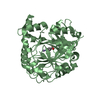
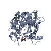
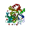
- PDB-3abf: Crystal Structure of a 4-Oxalocrotonate Tautomerase Homologue (TT... -
+ Open data

- Basic information
| Entry | Database: PDB / ID: 3abf | ||||||
|---|---|---|---|---|---|---|---|
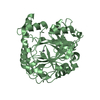
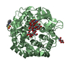
| Title | Crystal Structure of a 4-Oxalocrotonate Tautomerase Homologue (TTHB242) | ||||||
 Components Components | 4-oxalocrotonate tautomerase | ||||||
 Keywords Keywords | ISOMERASE / tautomerase | ||||||
| Function / homology | 4-oxalocrotonate tautomerase / Tautomerase enzyme / Macrophage Migration Inhibitory Factor / Macrophage Migration Inhibitory Factor / Tautomerase/MIF superfamily / isomerase activity / 2-Layer Sandwich / Alpha Beta / 4-oxalocrotonate tautomerase Function and homology information Function and homology information | ||||||
| Biological species |   Thermus thermophilus (bacteria) Thermus thermophilus (bacteria) | ||||||
| Method |  X-RAY DIFFRACTION / SYNCHROTRON / MOLECULAR REPLACEMENT / Resolution: 1.94 Å X-RAY DIFFRACTION / SYNCHROTRON / MOLECULAR REPLACEMENT / Resolution: 1.94 Å | ||||||
 Authors Authors | Kida, H. / Miki, K. | ||||||
 Citation Citation | Journal: To be Published Title: Crystal Structure of a 4-Oxalocrotonate Tautomerase Homologue (TTHB242) from Thermus thermophilus HB8 Authors: Kida, H. / Kita, A. / Miki, K. | ||||||
| History |
|
- Structure visualization
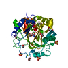
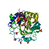
| Structure viewer | Molecule: MolmilJmol/JSmol |
|---|
- Downloads & links
-Download
| PDBx/mmCIF format | 3abf.cif.gz | 89.5 KB | Display | PDBx/mmCIF format |
|---|---|---|---|---|
| PDB format | pdb3abf.ent.gz | 68.6 KB | Display | PDB format |
| PDBx/mmJSON format | 3abf.json.gz | Tree view | PDBx/mmJSON format | |
| Others |  Other downloads Other downloads |
-Validation report
| Arichive directory | https://data.pdbj.org/pub/pdb/validation_reports/ab/3abfftp://data.pdbj.org/pub/pdb/validation_reports/ab/3abf | HTTPS FTP |
|---|
-Related structure data
| Related structure data |  1bjpS S: Starting model for refinement |
|---|---|
| Similar structure data |


-Links
 PDBj
PDBj- Assembly
| Deposited unit | 
| ||||||||
|---|---|---|---|---|---|---|---|---|---|
| 1 |
| ||||||||
| Unit cell |
|
-Components
| #1: Protein | Mass: 7426.663 Da / Num. of mol.: 6 Source method: isolated from a genetically manipulated source Source: (gene. exp.) Thermus thermophilus (bacteria) / Strain: HB8 / Gene: TTHB242 / Plasmid: pET11a / Production host: #2: Chemical |   Mass: 96.063 Da / Num. of mol.: 3 / Source method: obtained synthetically / Formula: SO4 Mass: 96.063 Da / Num. of mol.: 3 / Source method: obtained synthetically / Formula: SO4#3: Water | ChemComp-HOH / |  Mass: 18.015 Da / Num. of mol.: 195 / Source method: isolated from a natural source / Formula: H2O Mass: 18.015 Da / Num. of mol.: 195 / Source method: isolated from a natural source / Formula: H2O |
|---|
-Experimental details
-Experiment
| Experiment | Method: X-RAY DIFFRACTION / Number of used crystals: 1 |
|---|
- Sample preparation
| Crystal | Density Matthews: 1.97 Å3/Da / Density % sol: 37.53 % |
|---|---|
| Crystal grow | Temperature: 290 K / pH: 5.6 Details: 0.6M MgSO4, 0.1M sodium citrate pH 5.6, vapor diffusion, sitting drop, temperature 290K |
-Data collection
| Diffraction | Mean temperature: 293 K | ||||||||||||||||||||||||||||||||||||||||||||||||||||||||||||||||||
|---|---|---|---|---|---|---|---|---|---|---|---|---|---|---|---|---|---|---|---|---|---|---|---|---|---|---|---|---|---|---|---|---|---|---|---|---|---|---|---|---|---|---|---|---|---|---|---|---|---|---|---|---|---|---|---|---|---|---|---|---|---|---|---|---|---|---|---|
| Diffraction source | Source: SYNCHROTRON / Site: Photon Factory  / Beamline: BL-6A / Wavelength: 0.978 Å / Beamline: BL-6A / Wavelength: 0.978 Å | ||||||||||||||||||||||||||||||||||||||||||||||||||||||||||||||||||
| Detector | Type: ADSC QUANTUM 4r / Detector: CCD / Date: Oct 12, 2002 | ||||||||||||||||||||||||||||||||||||||||||||||||||||||||||||||||||
| Radiation | Protocol: SINGLE WAVELENGTH / Monochromatic (M) / Laue (L): M / Scattering type: x-ray | ||||||||||||||||||||||||||||||||||||||||||||||||||||||||||||||||||
| Radiation wavelength | Wavelength: 0.978 Å / Relative weight: 1 | ||||||||||||||||||||||||||||||||||||||||||||||||||||||||||||||||||
| Reflection | Resolution: 1.94→50 Å / Num. obs: 26856 / % possible obs: 99.7 % / Redundancy: 4 % / Rmerge(I) obs: 0.057 / Χ2: 1 / Net I/σ(I): 22.231 | ||||||||||||||||||||||||||||||||||||||||||||||||||||||||||||||||||
| Reflection shell |
|
- Processing
| Software |
| ||||||||||||||||||||||||||||||||||||||||||||||||||||||||||||||||||||||||||||||||
|---|---|---|---|---|---|---|---|---|---|---|---|---|---|---|---|---|---|---|---|---|---|---|---|---|---|---|---|---|---|---|---|---|---|---|---|---|---|---|---|---|---|---|---|---|---|---|---|---|---|---|---|---|---|---|---|---|---|---|---|---|---|---|---|---|---|---|---|---|---|---|---|---|---|---|---|---|---|---|---|---|---|
| Refinement | Method to determine structure: MOLECULAR REPLACEMENT Starting model: PDB ENTRY 1BJP Resolution: 1.94→43.52 Å / Occupancy max: 1 / Occupancy min: 0
| ||||||||||||||||||||||||||||||||||||||||||||||||||||||||||||||||||||||||||||||||
| Solvent computation | Bsol: 67.79 Å2 | ||||||||||||||||||||||||||||||||||||||||||||||||||||||||||||||||||||||||||||||||
| Displacement parameters | Biso mean: 33.33 Å2
| ||||||||||||||||||||||||||||||||||||||||||||||||||||||||||||||||||||||||||||||||
| Refinement step | Cycle: LAST / Resolution: 1.94→43.52 Å
| ||||||||||||||||||||||||||||||||||||||||||||||||||||||||||||||||||||||||||||||||
| Refine LS restraints |
| ||||||||||||||||||||||||||||||||||||||||||||||||||||||||||||||||||||||||||||||||
| Xplor file |
|
