 Movie
Movie Controller

[English] 日本語
 Yorodumi
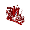
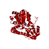
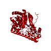
- PDB-3aat: ACTIVITY AND STRUCTURE OF THE ACTIVE-SITE MUTANTS R386Y AND R386F... -
+ Open data

- Basic information
| Entry | Database: PDB / ID: 3aat | ||||||
|---|---|---|---|---|---|---|---|
| Title | ACTIVITY AND STRUCTURE OF THE ACTIVE-SITE MUTANTS R386Y AND R386F OF ESCHERICHIA COLI ASPARTATE AMINOTRANSFERASE | ||||||
 Components Components | ASPARTATE AMINOTRANSFERASE | ||||||
 Keywords Keywords | TRANSFERASE(AMINOTRANSFERASE) | ||||||
| Function / homology |  Function and homology information Function and homology information: / L-tyrosine:2-oxoglutarate transaminase activity / L-phenylalanine biosynthetic process / aspartate transaminase / L-aspartate:2-oxoglutarate transaminase activity / pyridoxal phosphate binding / protein homodimerization activity / identical protein binding / cytosol / cytoplasm Similarity search - Function | ||||||
| Biological species |  | ||||||
| Method |  X-RAY DIFFRACTION / Resolution: 2.8 Å X-RAY DIFFRACTION / Resolution: 2.8 Å | ||||||
 Authors Authors | Danishefsky, A.T. / Ringe, D. / Petsko, G.A. | ||||||
 Citation Citation | Journal: Biochemistry / Year: 1991 Title: Activity and structure of the active-site mutants R386Y and R386F of Escherichia coli aspartate aminotransferase. Authors: Danishefsky, A.T. / Onnufer, J.J. / Petsko, G.A. / Ringe, D. #1: Journal: Biochemistry / Year: 1989Title: 2.8-Angstroms-Resolution Crystal Structure of an Active-Site Mutant of Aspartate Aminotransferse from Escherichia Coli Authors: Smith, D.L. / Almo, S.C. / Toney, M.D. / Ringe, D. #2: Journal: J.Mol.Biol. / Year: 1986Title: Preliminary X-Ray Data for Aspartate Aminotransferase from Escherichia Coli Authors: Smith, D.L. / Ringe, D. / Finlayson, W.L. / Kirsch, J.F. | ||||||
| History |
|
- Structure visualization
| Structure viewer | Molecule: MolmilJmol/JSmol |
|---|
- Downloads & links
-Download
| PDBx/mmCIF format | 3aat.cif.gz | 84.1 KB | Display | PDBx/mmCIF format |
|---|---|---|---|---|
| PDB format | pdb3aat.ent.gz | 58.7 KB | Display | PDB format |
| PDBx/mmJSON format | 3aat.json.gz | Tree view | PDBx/mmJSON format | |
| Others |  Other downloads Other downloads |
-Validation report
| Arichive directory | https://data.pdbj.org/pub/pdb/validation_reports/aa/3aatftp://data.pdbj.org/pub/pdb/validation_reports/aa/3aat | HTTPS FTP |
|---|
-Related structure data
| Similar structure data |
|---|

-Links
 PDBj
PDBj- Assembly
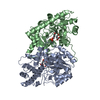
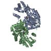
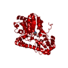
| Deposited unit | 
| ||||||||
|---|---|---|---|---|---|---|---|---|---|
| 1 | 
| ||||||||
| Unit cell |
| ||||||||
| Atom site foot note | 1: RESIDUES 138 AND 195 ARE CIS PROLINES. | ||||||||
| Details | THE MOLECULE IS A DIMER. TO GENERATE THE OTHER CHAIN ONE MUST APPLY THE CRYSTALLOGRAPHIC SYMMETRY OPERATION (X, 87.60-Y, 78.80-Z) TO THE COORDINATES IN THIS ENTRY. |
-Components
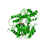
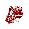
| #1: Protein | Mass: 43609.191 Da / Num. of mol.: 1 / Mutation: R374F Source method: isolated from a genetically manipulated source Source: (gene. exp.) |
|---|---|
| #2: Chemical | ChemComp-SO4 /   Mass: 96.063 Da / Num. of mol.: 1 / Source method: obtained synthetically / Formula: SO4 Mass: 96.063 Da / Num. of mol.: 1 / Source method: obtained synthetically / Formula: SO4 |
| #3: Chemical | ChemComp-PLP /   Mass: 247.142 Da / Num. of mol.: 1 / Source method: obtained synthetically / Formula: C8H10NO6P Mass: 247.142 Da / Num. of mol.: 1 / Source method: obtained synthetically / Formula: C8H10NO6P |
| Sequence details | THE RESIDUE NUMBERING USED IN THIS ENTRY HAS BEEN CHOSEN TO MAXIMIZE HOMOLOGY WITH OTHER SPECIES OF ...THE RESIDUE NUMBERING USED IN THIS ENTRY HAS BEEN CHOSEN TO MAXIMIZE HOMOLOGY WITH OTHER SPECIES OF THE ENZYME. THEREFORE, THE RESIDUE NUMBERING STARTS WITH 5 AND RESIDUE NUMBERS 128 - 132, 153, 232, AND 407 HAVE NOT BEEN USED. |
-Experimental details
-Experiment
| Experiment | Method: X-RAY DIFFRACTION |
|---|
- Sample preparation
| Crystal | Density Matthews: 3.09 Å3/Da / Density % sol: 60.14 % | |||||||||||||||
|---|---|---|---|---|---|---|---|---|---|---|---|---|---|---|---|---|
| Crystal grow | *PLUS Method: vapor diffusion | |||||||||||||||
| Components of the solutions | *PLUS
|
-Data collection
| Radiation | Scattering type: x-ray |
|---|---|
| Radiation wavelength | Relative weight: 1 |
| Reflection | *PLUS Highest resolution: 2.5 Å / Rmerge(I) obs: 0.067 |
- Processing
| Software | Name: PROLSQ / Classification: refinement | ||||||||||||||||||||||||||||||||||||||||||||||||||||||||||||||||||||||||||||||||||||
|---|---|---|---|---|---|---|---|---|---|---|---|---|---|---|---|---|---|---|---|---|---|---|---|---|---|---|---|---|---|---|---|---|---|---|---|---|---|---|---|---|---|---|---|---|---|---|---|---|---|---|---|---|---|---|---|---|---|---|---|---|---|---|---|---|---|---|---|---|---|---|---|---|---|---|---|---|---|---|---|---|---|---|---|---|---|
| Refinement | Resolution: 2.8→10 Å / Rfactor obs: 0.215 Details: THE GEOMETRY AT MANY OF THE COIL REGIONS WAS NOT CLEARLY DEFINED IN THE ELECTRON DENSITY MAPS. | ||||||||||||||||||||||||||||||||||||||||||||||||||||||||||||||||||||||||||||||||||||
| Refinement step | Cycle: LAST / Resolution: 2.8→10 Å
| ||||||||||||||||||||||||||||||||||||||||||||||||||||||||||||||||||||||||||||||||||||
| Refine LS restraints |
| ||||||||||||||||||||||||||||||||||||||||||||||||||||||||||||||||||||||||||||||||||||
| Refinement | *PLUS Highest resolution: 2.5 Å / Lowest resolution: 10 Å / Num. reflection obs: 10531 / σ(I): 1 / Rfactor obs: 0.215 | ||||||||||||||||||||||||||||||||||||||||||||||||||||||||||||||||||||||||||||||||||||
| Solvent computation | *PLUS | ||||||||||||||||||||||||||||||||||||||||||||||||||||||||||||||||||||||||||||||||||||
| Displacement parameters | *PLUS Biso mean: 12 Å2 |
