 Movie
Movie Controller


+ Open data

- Basic information
| Entry | Database: PDB / ID: 3a5i | ||||||
|---|---|---|---|---|---|---|---|
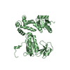
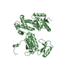
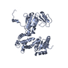
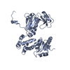
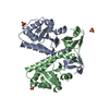
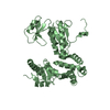
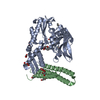
| Title | Structure of the cytoplasmic domain of FlhA | ||||||
 Components Components | Flagellar biosynthesis protein flhA | ||||||
 Keywords Keywords | PROTEIN TRANSPORT / four domains / thioredoxin-like fold / Bacterial flagellum biogenesis / Bacterial flagellum protein export / Cell inner membrane / Cell membrane / Membrane / Transmembrane / Transport | ||||||
| Function / homology |  Function and homology information Function and homology information | ||||||
| Biological species |  Salmonella typhimurium (bacteria) Salmonella typhimurium (bacteria) | ||||||
| Method |  X-RAY DIFFRACTION / SYNCHROTRON / MAD / Resolution: 2.8 Å X-RAY DIFFRACTION / SYNCHROTRON / MAD / Resolution: 2.8 Å | ||||||
 Authors Authors | Imada, K. / Saijo-Hamano, Y. / Shimada, M. / Namba, K. | ||||||
 Citation Citation | Journal: Mol.Microbiol. / Year: 2010 Title: Structure of the cytoplasmic domain of FlhA and implication for flagellar type III protein export Authors: Saijo-Hamano, Y. / Imada, K. / Minamino, T. / Kihara, M. / Shimada, M. / Kitao, A. / Namba, K. #1: Journal: Acta Crystallogr.,Sect.F / Year: 2005 Title: Crystallization and preliminary X-ray analysis of the C-terminal cytoplasmic domain of FlhA, a membrane-protein subunit of the bacterial flagellar type III protein-export apparatus Authors: Saijo-Hamano, Y. / Imada, K. / Minamino, T. / Kihara, M. / Macnab, R.M. / Namba, K. | ||||||
| History |
|
- Structure visualization
| Structure viewer | Molecule: MolmilJmol/JSmol |
|---|
- Downloads & links
-Download
| PDBx/mmCIF format | 3a5i.cif.gz | 141.8 KB | Display | PDBx/mmCIF format |
|---|---|---|---|---|
| PDB format | pdb3a5i.ent.gz | 112.5 KB | Display | PDB format |
| PDBx/mmJSON format | 3a5i.json.gz | Tree view | PDBx/mmJSON format | |
| Others |  Other downloads Other downloads |
-Validation report
| Summary document | 3a5i_validation.pdf.gz | 446.8 KB | Display | wwPDB validaton report |
|---|---|---|---|---|
| Full document | 3a5i_full_validation.pdf.gz | 479.9 KB | Display | |
| Data in XML | 3a5i_validation.xml.gz | 28.7 KB | Display | |
| Data in CIF | 3a5i_validation.cif.gz | 38.3 KB | Display | |
| Arichive directory | https://data.pdbj.org/pub/pdb/validation_reports/a5/3a5iftp://data.pdbj.org/pub/pdb/validation_reports/a5/3a5i | HTTPS FTP |
-Related structure data
| Similar structure data |
|---|


-Links
 PDBj
PDBj- Assembly
| Deposited unit | 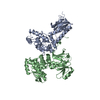
| ||||||||
|---|---|---|---|---|---|---|---|---|---|
| 1 | 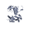
| ||||||||
| 2 | 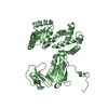
| ||||||||
| Unit cell |
| ||||||||
| Details | AUTHOR DETERMINED BIOLOGICAL UNIT: UNKNOWN |
-Components
| #1: Protein | Mass: 43258.348 Da / Num. of mol.: 2 / Fragment: FlhAc, UNP residues 328-692 Source method: isolated from a genetically manipulated source Source: (gene. exp.) Salmonella typhimurium (bacteria) / Strain: SJW1103 / Gene: flhA, STM1913 / Plasmid: pET19b / Production host: |
|---|
-Experimental details
-Experiment
| Experiment | Method: X-RAY DIFFRACTION / Number of used crystals: 2 |
|---|
- Sample preparation
| Crystal |
| |||||||||
|---|---|---|---|---|---|---|---|---|---|---|
| Crystal grow | Temperature: 293 K / Method: vapor diffusion, sitting drop / pH: 8.5 Details: 10-15% PEG300, 4-6% PEG8000, 0.1M Tris-HCl, 0.5mM EDTA, 10-15% glycerol, pH8.5, VAPOR DIFFUSION, SITTING DROP, temperature 293K |
-Data collection
| Diffraction |
| ||||||||||||||||||
|---|---|---|---|---|---|---|---|---|---|---|---|---|---|---|---|---|---|---|---|
| Diffraction source |
| ||||||||||||||||||
| Detector |
| ||||||||||||||||||
| Radiation |
| ||||||||||||||||||
| Radiation wavelength |
| ||||||||||||||||||
| Reflection | Resolution: 2.8→40.8 Å / Num. all: 37317 / Num. obs: 37317 / % possible obs: 99.8 % / Observed criterion σ(F): 0 / Observed criterion σ(I): 0 / Redundancy: 4.9 % / Biso Wilson estimate: 85.2 Å2 / Rmerge(I) obs: 0.084 / Net I/σ(I): 13.8 | ||||||||||||||||||
| Reflection shell | Resolution: 2.8→2.95 Å / Redundancy: 4.7 % / Rmerge(I) obs: 0.398 / Mean I/σ(I) obs: 3 / Num. unique all: 5343 / % possible all: 98.8 |

- Processing
| Software |
| ||||||||||||||||||||||||||||||||||||
|---|---|---|---|---|---|---|---|---|---|---|---|---|---|---|---|---|---|---|---|---|---|---|---|---|---|---|---|---|---|---|---|---|---|---|---|---|---|
| Refinement | Method to determine structure: MAD / Resolution: 2.8→40.8 Å / Rfactor Rfree error: 0.007 / Data cutoff high absF: 2106476.83 / Data cutoff low absF: 0 / Isotropic thermal model: RESTRAINED / Cross valid method: THROUGHOUT / σ(F): 0 / Stereochemistry target values: Engh & Huber
| ||||||||||||||||||||||||||||||||||||
| Solvent computation | Solvent model: FLAT MODEL / Bsol: 29.0094 Å2 / ksol: 0.29557 e/Å3 | ||||||||||||||||||||||||||||||||||||
| Displacement parameters | Biso mean: 75.2 Å2
| ||||||||||||||||||||||||||||||||||||
| Refine analyze |
| ||||||||||||||||||||||||||||||||||||
| Refinement step | Cycle: LAST / Resolution: 2.8→40.8 Å
| ||||||||||||||||||||||||||||||||||||
| Refine LS restraints |
| ||||||||||||||||||||||||||||||||||||
| LS refinement shell | Resolution: 2.8→2.98 Å / Rfactor Rfree error: 0.025 / Total num. of bins used: 6
| ||||||||||||||||||||||||||||||||||||
| Xplor file |
|
