+ Open data
- Basic information
| Entry | Database: PDB / ID: 3a0h | |||||||||
|---|---|---|---|---|---|---|---|---|---|---|
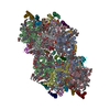
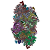
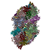
| Title | Crystal structure of I-substituted Photosystem II complex | |||||||||
 Components Components |
| |||||||||
 Keywords Keywords | ELECTRON TRANSPORT / MULTI-MEMBRANE PROTEIN COMPLEX / Herbicide resistance / Iron / Membrane / Metal-binding / Photosynthesis / Photosystem II / Thylakoid / Transmembrane / Transport / Heme / Reaction center | |||||||||
| Function / homology |  Function and homology information Function and homology informationphotosystem II oxygen evolving complex / photosystem II assembly / oxygen evolving activity / photosystem II stabilization / photosystem II reaction center / photosystem II / oxidoreductase activity, acting on diphenols and related substances as donors, oxygen as acceptor / photosynthetic electron transport chain / photosystem II / response to herbicide ...photosystem II oxygen evolving complex / photosystem II assembly / oxygen evolving activity / photosystem II stabilization / photosystem II reaction center / photosystem II / oxidoreductase activity, acting on diphenols and related substances as donors, oxygen as acceptor / photosynthetic electron transport chain / photosystem II / response to herbicide / extrinsic component of membrane / : / plasma membrane-derived thylakoid membrane / chlorophyll binding / photosynthetic electron transport in photosystem II / phosphate ion binding / photosynthesis, light reaction / photosynthesis / respiratory electron transport chain / electron transfer activity / protein stabilization / iron ion binding / heme binding / metal ion binding Similarity search - Function | |||||||||
| Biological species |  Thermosynechococcus vulcanus (bacteria) Thermosynechococcus vulcanus (bacteria) | |||||||||
| Method |  X-RAY DIFFRACTION / X-RAY DIFFRACTION /  SYNCHROTRON / SYNCHROTRON /  MOLECULAR REPLACEMENT / Resolution: 4 Å MOLECULAR REPLACEMENT / Resolution: 4 Å | |||||||||
 Authors Authors | Kawakami, K. / Umena, Y. / Kamiya, N. / Shen, J.-R. | |||||||||
 Citation Citation |  Journal: Proc.Natl.Acad.Sci.USA / Year: 2009 Journal: Proc.Natl.Acad.Sci.USA / Year: 2009Title: Location of chloride and its possible functions in oxygen-evolving photosystem II revealed by X-ray crystallography Authors: Kawakami, K. / Umena, Y. / Kamiya, N. / Shen, J.-R. | |||||||||
| History |
|
- Structure visualization
| Structure viewer | Molecule:  Molmil Molmil Jmol/JSmol Jmol/JSmol |
|---|
- Downloads & links
- Download
| PDBx/mmCIF format |  3a0h.cif.gz 3a0h.cif.gz | 1.2 MB | Display |  PDBx/mmCIF format PDBx/mmCIF format |
|---|---|---|---|---|
| PDB format |  pdb3a0h.ent.gz pdb3a0h.ent.gz | 1001.7 KB | Display |  PDB format PDB format |
| PDBx/mmJSON format |  3a0h.json.gz 3a0h.json.gz | Tree view |  PDBx/mmJSON format PDBx/mmJSON format | |
| Others |  Other downloads Other downloads |
-Validation report
| Arichive directory |  https://data.pdbj.org/pub/pdb/validation_reports/a0/3a0h https://data.pdbj.org/pub/pdb/validation_reports/a0/3a0h ftp://data.pdbj.org/pub/pdb/validation_reports/a0/3a0h ftp://data.pdbj.org/pub/pdb/validation_reports/a0/3a0h | HTTPS FTP |
|---|
-Related structure data
| Related structure data | 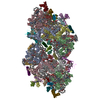 3a0bC 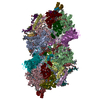 2axtS C: citing same article ( S: Starting model for refinement |
|---|---|
| Similar structure data |
- Links
Links
- Assembly
| Deposited unit | 
| ||||||||
|---|---|---|---|---|---|---|---|---|---|
| 1 |
| ||||||||
| Unit cell |
|
- Components
-Protein , 2 types, 4 molecules AaVv
| #1: Protein | Mass: 38295.684 Da / Num. of mol.: 2 / Source method: isolated from a natural source / Source: (natural)  Thermosynechococcus vulcanus (bacteria) / Cellular location: thylakoid membrane / References: UniProt: P51765 Thermosynechococcus vulcanus (bacteria) / Cellular location: thylakoid membrane / References: UniProt: P51765#16: Protein | Mass: 15148.255 Da / Num. of mol.: 2 / Source method: isolated from a natural source / Source: (natural)  Thermosynechococcus vulcanus (bacteria) / Cellular location: thylakoid membrane / References: UniProt: P0A387 Thermosynechococcus vulcanus (bacteria) / Cellular location: thylakoid membrane / References: UniProt: P0A387 |
|---|
-Photosystem II ... , 16 types, 32 molecules BbCcDdHhIiJjKkLlMmOoTtUuXxYyNnZz
| #2: Protein | Mass: 54042.477 Da / Num. of mol.: 2 / Source method: isolated from a natural source / Source: (natural)  Thermosynechococcus vulcanus (bacteria) / Cellular location: thylakoid membrane / References: UniProt: D0VWR1*PLUS Thermosynechococcus vulcanus (bacteria) / Cellular location: thylakoid membrane / References: UniProt: D0VWR1*PLUS#3: Protein | Mass: 48763.777 Da / Num. of mol.: 2 / Source method: isolated from a natural source / Source: (natural)  Thermosynechococcus vulcanus (bacteria) / Cellular location: thylakoid membrane / References: UniProt: D0VWR7*PLUS Thermosynechococcus vulcanus (bacteria) / Cellular location: thylakoid membrane / References: UniProt: D0VWR7*PLUS#4: Protein | Mass: 38118.641 Da / Num. of mol.: 2 / Source method: isolated from a natural source / Source: (natural)  Thermosynechococcus vulcanus (bacteria) / Cellular location: thylakoid membrane / References: UniProt: D0VWR8*PLUS Thermosynechococcus vulcanus (bacteria) / Cellular location: thylakoid membrane / References: UniProt: D0VWR8*PLUS#7: Protein | Mass: 7170.506 Da / Num. of mol.: 2 / Source method: isolated from a natural source / Source: (natural)  Thermosynechococcus vulcanus (bacteria) / Cellular location: thylakoid membrane / References: UniProt: P19052*PLUS Thermosynechococcus vulcanus (bacteria) / Cellular location: thylakoid membrane / References: UniProt: P19052*PLUS#8: Protein/peptide | Mass: 4052.885 Da / Num. of mol.: 2 / Source method: isolated from a natural source / Source: (natural)  Thermosynechococcus vulcanus (bacteria) / Cellular location: thylakoid membrane / References: UniProt: P12240*PLUS Thermosynechococcus vulcanus (bacteria) / Cellular location: thylakoid membrane / References: UniProt: P12240*PLUS#9: Protein/peptide | Mass: 4105.908 Da / Num. of mol.: 2 / Source method: isolated from a natural source / Source: (natural)  Thermosynechococcus vulcanus (bacteria) / Cellular location: thylakoid membrane / References: UniProt: Q7DGD4 Thermosynechococcus vulcanus (bacteria) / Cellular location: thylakoid membrane / References: UniProt: Q7DGD4#10: Protein/peptide | Mass: 3891.654 Da / Num. of mol.: 2 / Source method: isolated from a natural source / Source: (natural)  Thermosynechococcus vulcanus (bacteria) / Cellular location: thylakoid membrane / References: UniProt: P19054*PLUS Thermosynechococcus vulcanus (bacteria) / Cellular location: thylakoid membrane / References: UniProt: P19054*PLUS#11: Protein/peptide | Mass: 4299.044 Da / Num. of mol.: 2 / Source method: isolated from a natural source / Source: (natural)  Thermosynechococcus vulcanus (bacteria) / Cellular location: thylakoid membrane / References: UniProt: P12241 Thermosynechococcus vulcanus (bacteria) / Cellular location: thylakoid membrane / References: UniProt: P12241#12: Protein/peptide | Mass: 4015.688 Da / Num. of mol.: 2 / Source method: isolated from a natural source / Source: (natural)  Thermosynechococcus vulcanus (bacteria) / Cellular location: thylakoid membrane / References: UniProt: P12312*PLUS Thermosynechococcus vulcanus (bacteria) / Cellular location: thylakoid membrane / References: UniProt: P12312*PLUS#13: Protein | Mass: 26452.498 Da / Num. of mol.: 2 / Source method: isolated from a natural source / Source: (natural)  Thermosynechococcus vulcanus (bacteria) / Cellular location: thylakoid membrane / References: UniProt: D0VWR2*PLUS Thermosynechococcus vulcanus (bacteria) / Cellular location: thylakoid membrane / References: UniProt: D0VWR2*PLUS#14: Protein/peptide | Mass: 3620.369 Da / Num. of mol.: 2 / Source method: isolated from a natural source / Source: (natural)  Thermosynechococcus vulcanus (bacteria) / Cellular location: thylakoid membrane / References: UniProt: P12313 Thermosynechococcus vulcanus (bacteria) / Cellular location: thylakoid membrane / References: UniProt: P12313#15: Protein | Mass: 11095.432 Da / Num. of mol.: 2 / Source method: isolated from a natural source / Source: (natural)  Thermosynechococcus vulcanus (bacteria) / Cellular location: thylakoid membrane / References: UniProt: P56152*PLUS Thermosynechococcus vulcanus (bacteria) / Cellular location: thylakoid membrane / References: UniProt: P56152*PLUS#17: Protein/peptide | Mass: 3477.162 Da / Num. of mol.: 2 / Source method: isolated from a natural source / Source: (natural)  Thermosynechococcus vulcanus (bacteria) / Cellular location: thylakoid membrane / References: UniProt: D0VWR4*PLUS Thermosynechococcus vulcanus (bacteria) / Cellular location: thylakoid membrane / References: UniProt: D0VWR4*PLUS#18: Protein/peptide | Mass: 2999.790 Da / Num. of mol.: 2 / Source method: isolated from a natural source / Source: (natural)  Thermosynechococcus vulcanus (bacteria) / Cellular location: thylakoid membrane / References: UniProt: D0VWR3*PLUS Thermosynechococcus vulcanus (bacteria) / Cellular location: thylakoid membrane / References: UniProt: D0VWR3*PLUS#19: Protein/peptide | Mass: 1975.426 Da / Num. of mol.: 2 / Source method: isolated from a natural source / Source: (natural)  Thermosynechococcus vulcanus (bacteria) / Cellular location: thylakoid membrane Thermosynechococcus vulcanus (bacteria) / Cellular location: thylakoid membrane#20: Protein | Mass: 6766.187 Da / Num. of mol.: 2 / Source method: isolated from a natural source / Source: (natural)  Thermosynechococcus vulcanus (bacteria) / Cellular location: thylakoid membrane / References: UniProt: D0VWR5*PLUS Thermosynechococcus vulcanus (bacteria) / Cellular location: thylakoid membrane / References: UniProt: D0VWR5*PLUS |
|---|
-Cytochrome b559 subunit ... , 2 types, 4 molecules EeFf
| #5: Protein | Mass: 9477.697 Da / Num. of mol.: 2 / Source method: isolated from a natural source / Source: (natural)  Thermosynechococcus vulcanus (bacteria) / Cellular location: thylakoid membrane / References: UniProt: P12238 Thermosynechococcus vulcanus (bacteria) / Cellular location: thylakoid membrane / References: UniProt: P12238#6: Protein/peptide | Mass: 4936.704 Da / Num. of mol.: 2 / Source method: isolated from a natural source / Source: (natural)  Thermosynechococcus vulcanus (bacteria) / Cellular location: thylakoid membrane / References: UniProt: P12239 Thermosynechococcus vulcanus (bacteria) / Cellular location: thylakoid membrane / References: UniProt: P12239 |
|---|
-Sugars , 1 types, 8 molecules 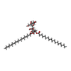
| #29: Sugar | ChemComp-DGD / |
|---|
-Non-polymers , 10 types, 128 molecules 
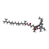

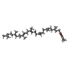
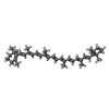


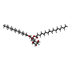


| #21: Chemical | | #22: Chemical | ChemComp-CLA / #23: Chemical | ChemComp-PHO / #24: Chemical | ChemComp-PQ9 / #25: Chemical | ChemComp-BCR / #26: Chemical | #27: Chemical | ChemComp-IOD / #28: Chemical | ChemComp-MGE / ( #30: Chemical | #31: Chemical | ChemComp-HEM / |
|---|
-Details
| Compound details | THIS ENTRY IS PHOTOSYSTE| Has protein modification | Y | Nonpolymer details | CHLORIDE IONS LOCATED IN THE OXYGEN-EVOLVING CENTER WERE SUBSTITUTE | Sequence details | THERE IS NO DATABASE REFERENCE FOR CHAINS B,C,D,H,K,M,O,U,Y, X, AND Z AT THE TIME OF PROCESSING. ...THERE IS NO DATABASE REFERENCE FOR CHAINS B,C,D,H,K,M,O,U,Y, X, AND Z AT THE TIME OF PROCESSING | |
|---|
-Experimental details
-Experiment
| Experiment | Method:  X-RAY DIFFRACTION / Number of used crystals: 1 X-RAY DIFFRACTION / Number of used crystals: 1 |
|---|
- Sample preparation
| Crystal | Density Matthews: 3.76 Å3/Da / Density % sol: 67.29 % |
|---|---|
| Crystal grow | Temperature: 293 K / Method: vapor diffusion, hanging drop / pH: 6 Details: 4% PEG 1450, 40mM MgSO4, 10mM MgCl2, 13% glycerol, 0.01% DM, 20mM MES (pH6.0), 10mM NaI, 5mM CaI, VAPOR DIFFUSION, HANGING DROP, temperature 293K |
-Data collection
| Diffraction | Mean temperature: 100 K |
|---|---|
| Diffraction source | Source:  SYNCHROTRON / Site: SYNCHROTRON / Site:  SPring-8 SPring-8  / Beamline: BL41XU / Wavelength: 1 Å / Beamline: BL41XU / Wavelength: 1 Å |
| Detector | Type: ADSC QUANTUM 315 / Detector: CCD / Date: Jun 30, 2007 |
| Radiation | Monochromator: rotated-inclined double-crystal / Protocol: SINGLE WAVELENGTH / Monochromatic (M) / Laue (L): M / Scattering type: x-ray |
| Radiation wavelength | Wavelength: 1 Å / Relative weight: 1 |
| Reflection | Resolution: 4→50 Å / Num. all: 75277 / Num. obs: 75277 / % possible obs: 99.8 % / Observed criterion σ(F): 0 / Observed criterion σ(I): -3 / Redundancy: 7.3 % / Rmerge(I) obs: 0.09 / Rsym value: 0.09 / Net I/σ(I): 25.57 |
| Reflection shell | Resolution: 4→4.14 Å / Redundancy: 7.4 % / Rmerge(I) obs: 0.908 / Mean I/σ(I) obs: 2.158 / Num. unique all: 7447 / Rsym value: 0.908 / % possible all: 100 |
- Processing
| Software |
| ||||||||||||||||||||||||||||||||||||||||||||||||||||||||||||||||||||||||||||||||
|---|---|---|---|---|---|---|---|---|---|---|---|---|---|---|---|---|---|---|---|---|---|---|---|---|---|---|---|---|---|---|---|---|---|---|---|---|---|---|---|---|---|---|---|---|---|---|---|---|---|---|---|---|---|---|---|---|---|---|---|---|---|---|---|---|---|---|---|---|---|---|---|---|---|---|---|---|---|---|---|---|---|
| Refinement | Method to determine structure:  MOLECULAR REPLACEMENT MOLECULAR REPLACEMENTStarting model: PDB ENTRY 2AXT Resolution: 4→20 Å / Rfactor Rfree error: 0.005 / Data cutoff high absF: 7949177.06 / Data cutoff low absF: 0 / Isotropic thermal model: RESTRAINED / Cross valid method: THROUGHOUT / σ(F): 0 / σ(I): -3 / Stereochemistry target values: Engh & Huber / Details: BULK SOLVENT MODEL USED
| ||||||||||||||||||||||||||||||||||||||||||||||||||||||||||||||||||||||||||||||||
| Solvent computation | Solvent model: FLAT MODEL / Bsol: 84.842 Å2 / ksol: 0.25 e/Å3 | ||||||||||||||||||||||||||||||||||||||||||||||||||||||||||||||||||||||||||||||||
| Displacement parameters | Biso mean: 164.4 Å2
| ||||||||||||||||||||||||||||||||||||||||||||||||||||||||||||||||||||||||||||||||
| Refine analyze |
| ||||||||||||||||||||||||||||||||||||||||||||||||||||||||||||||||||||||||||||||||
| Refinement step | Cycle: LAST / Resolution: 4→20 Å
| ||||||||||||||||||||||||||||||||||||||||||||||||||||||||||||||||||||||||||||||||
| Refine LS restraints |
| ||||||||||||||||||||||||||||||||||||||||||||||||||||||||||||||||||||||||||||||||
| LS refinement shell | Resolution: 4→4.25 Å / Rfactor Rfree error: 0.018 / Total num. of bins used: 6
| ||||||||||||||||||||||||||||||||||||||||||||||||||||||||||||||||||||||||||||||||
| Xplor file |
|
 Movie
Movie Controller






 PDBj
PDBj


