 Movie
Movie Controller

[English] 日本語
 Yorodumi
- PDB-38td: Cryo EM structure of a formate acetyltransferase (PFL) from Fanny... -
+ Open data

- Basic information
| Entry | Database: PDB / ID: 38td | |||||||||
|---|---|---|---|---|---|---|---|---|---|---|
| Title | Cryo EM structure of a formate acetyltransferase (PFL) from Fannyhessea vaginae in complex with CoA | |||||||||
 Components Components | Formate acetyltransferase | |||||||||
 Keywords Keywords | LYASE / SSGCID / STRUCTURAL GENOMICS / SEATTLE STRUCTURAL GENOMICS CENTER FOR INFECTIOUS DISEASE / formate acetyltransferase (PFL) / Fannyhessea vaginae | |||||||||
| Function / homology |  Function and homology information Function and homology informationformate C-acetyltransferase / formate C-acetyltransferase activity / glucose metabolic process / cytosol Similarity search - Function | |||||||||
| Biological species |  Fannyhessea vaginae DSM 15829 (bacteria) Fannyhessea vaginae DSM 15829 (bacteria) | |||||||||
| Method | ELECTRON MICROSCOPY / single particle reconstruction / cryo EM / Resolution: 2.96 Å | |||||||||
 Authors Authors | Liu, L. / Lovell, S. / Hammons, A.M. / Seattle Structural Genomics Center for Infectious Disease (SSGCID) | |||||||||
| Funding support |  United States, 2items United States, 2items
| |||||||||
 Citation Citation | Journal: To be published Title: Cryo EM structure of a formate acetyltransferase (PFL) from Fannyhessea vaginae in complex with CoA Authors: Liu, L. / Lovell, S. / Hammons, A.M. | |||||||||
| History |
|
- Structure visualization
| Structure viewer | Molecule: MolmilJmol/JSmol |
|---|
- Downloads & links
-Download
| PDBx/mmCIF format | 38td.cif.gz | 486.3 KB | Display | PDBx/mmCIF format |
|---|---|---|---|---|
| PDB format | pdb38td.ent.gz | 398 KB | Display | PDB format |
| PDBx/mmJSON format | 38td.json.gz | Tree view | PDBx/mmJSON format | |
| Others |  Other downloads Other downloads |
-Validation report
| Arichive directory | https://data.pdbj.org/pub/pdb/validation_reports/8t/38tdftp://data.pdbj.org/pub/pdb/validation_reports/8t/38td | HTTPS FTP |
|---|
-Related structure data
| Related structure data |  79135MC M: map data used to model this data C: citing same article ( |
|---|---|
| Similar structure data |
-Links
 PDBj
PDBj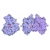

- Assembly
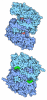
| Deposited unit | 
|
|---|---|
| 1 |
|
-Components
| #1: Protein | Mass: 83473.141 Da / Num. of mol.: 4 Source method: isolated from a genetically manipulated source Source: (gene. exp.) Fannyhessea vaginae DSM 15829 (bacteria)Gene: pflB, HMPREF0091_10019 / Plasmid: FavaA.20507.a.A1 / Production host:  Escherichia coli BL21(DE3) (bacteria) / References: UniProt: F1T4Y4, formate C-acetyltransferase Escherichia coli BL21(DE3) (bacteria) / References: UniProt: F1T4Y4, formate C-acetyltransferase#2: Chemical | ChemComp-COA / 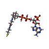 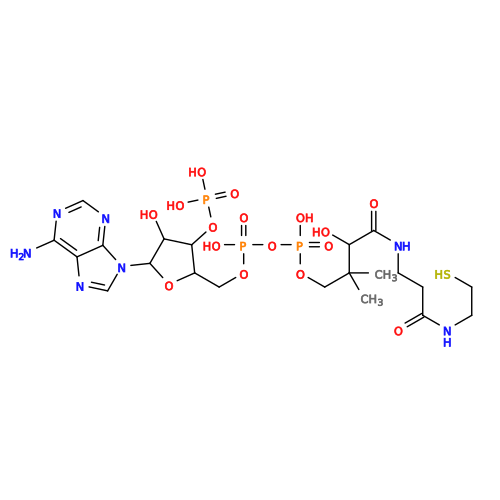 Mass: 767.534 Da / Num. of mol.: 4 / Source method: obtained synthetically / Formula: C21H36N7O16P3S / Feature type: SUBJECT OF INVESTIGATION Mass: 767.534 Da / Num. of mol.: 4 / Source method: obtained synthetically / Formula: C21H36N7O16P3S / Feature type: SUBJECT OF INVESTIGATIONHas ligand of interest | Y | Has protein modification | N | |
|---|
-Experimental details
-Experiment
| Experiment | Method: ELECTRON MICROSCOPY |
|---|---|
| EM experiment | Aggregation state: 2D ARRAY / 3D reconstruction method: single particle reconstruction |
- Sample preparation
| Component | Name: tetramer of formate acetyltransferase (PFL) / Type: COMPLEX / Entity ID: #1 / Source: RECOMBINANT |
|---|---|
| Molecular weight | Value: .33389 MDa / Experimental value: YES |
| Source (natural) | Organism: Fannyhessea vaginae DSM 15829 (bacteria) |
| Source (recombinant) | Organism: Escherichia coli (E. coli) / Strain: BL21(DE3) / Plasmid: FavaA.20507.a.A1 |
| Buffer solution | pH: 7 Details: 25 mM HEPES pH 7.0, 500 mM NaCl, 5% Glycerol, 2 mM DTT, 0.025% Azide |
| Specimen | Conc.: 0.5 mg/ml / Embedding applied: NO / Shadowing applied: NO / Staining applied: NO / Vitrification applied: YES |
| Specimen support | Grid material: COPPER / Grid mesh size: 300 divisions/in. / Grid type: Quantifoil R1.2/1.3 |
| Vitrification | Instrument: FEI VITROBOT MARK IV / Cryogen name: ETHANE / Humidity: 90 % / Chamber temperature: 277 K |
- Electron microscopy imaging
| Microscopy | Model: TFS GLACIOS |
|---|---|
| Electron gun | Electron source:  FIELD EMISSION GUN / Accelerating voltage: 200 kV / Illumination mode: FLOOD BEAM FIELD EMISSION GUN / Accelerating voltage: 200 kV / Illumination mode: FLOOD BEAM |
| Electron lens | Mode: 4D-STEM / Nominal magnification: 100000 X / Nominal defocus max: 4000 nm / Nominal defocus min: 100 nm / Cs: 2.7 mm / Alignment procedure: BASIC |
| Specimen holder | Cryogen: NITROGEN / Specimen holder model: FEI TITAN KRIOS AUTOGRID HOLDER |
| Image recording | Electron dose: 60 e/Å2 / Detector mode: COUNTING / Film or detector model: FEI FALCON I (4k x 4k) / Num. of grids imaged: 1 / Num. of real images: 5872 |
| EM imaging optics | Energyfilter name: TFS Selectris / Energyfilter slit width: 10 eV |
| Image scans | Width: 4096 / Height: 4096 |
- Processing
| EM software |
| ||||||||||||||||||||||||||||||||||||||||
|---|---|---|---|---|---|---|---|---|---|---|---|---|---|---|---|---|---|---|---|---|---|---|---|---|---|---|---|---|---|---|---|---|---|---|---|---|---|---|---|---|---|
| Image processing | Details: The selected images were high-pass filtered and normalized | ||||||||||||||||||||||||||||||||||||||||
| CTF correction | Type: PHASE FLIPPING AND AMPLITUDE CORRECTION | ||||||||||||||||||||||||||||||||||||||||
| Symmetry | Point symmetry: D2 (2x2 fold dihedral) | ||||||||||||||||||||||||||||||||||||||||
| 3D reconstruction | Resolution: 2.96 Å / Resolution method: FSC 0.143 CUT-OFF / Num. of particles: 2295925 / Symmetry type: 3D CRYSTAL | ||||||||||||||||||||||||||||||||||||||||
| Atomic model building | Protocol: FLEXIBLE FIT / Space: REAL | ||||||||||||||||||||||||||||||||||||||||
| Refinement | Highest resolution: 2.96 Å Stereochemistry target values: REAL-SPACE (WEIGHTED MAP SUM AT ATOM CENTERS) | ||||||||||||||||||||||||||||||||||||||||
| Refine LS restraints |
|
