 Movie
Movie Controller


+ Open data

- Basic information
| Entry | Database: PDB / ID: 38kl | |||||||||
|---|---|---|---|---|---|---|---|---|---|---|
| Title | Two AIP1 bound to the Barbed End of cofilin actin | |||||||||
 Components Components |
| |||||||||
 Keywords Keywords | STRUCTURAL PROTEIN / actin / cofilin / AIP1 | |||||||||
| Function / homology |  Function and homology information Function and homology informationmuscle thin filament assembly / positive regulation of actin filament depolymerization / negative regulation of actin filament polymerization / regulation of locomotion / actin filament depolymerization / locomotion / sarcomere organization / cytoskeletal motor activator activity / cortical actin cytoskeleton / myosin heavy chain binding ...muscle thin filament assembly / positive regulation of actin filament depolymerization / negative regulation of actin filament polymerization / regulation of locomotion / actin filament depolymerization / locomotion / sarcomere organization / cytoskeletal motor activator activity / cortical actin cytoskeleton / myosin heavy chain binding / tropomyosin binding / actin filament bundle / troponin I binding / filamentous actin / mesenchyme migration / sarcoplasm / myofibril / skeletal muscle myofibril / striated muscle thin filament / skeletal muscle thin filament assembly / actin filament bundle assembly / actin monomer binding / skeletal muscle fiber development / actin filament polymerization / stress fiber / titin binding / filopodium / actin filament / Hydrolases; Acting on acid anhydrides; Acting on acid anhydrides to facilitate cellular and subcellular movement / calcium-dependent protein binding / actin filament binding / lamellipodium / actin binding / cell body / protein domain specific binding / hydrolase activity / positive regulation of gene expression / calcium ion binding / magnesium ion binding / ATP binding / identical protein binding / cytoplasm Similarity search - Function | |||||||||
| Biological species |   Caenorhabditis elegans (invertebrata) Caenorhabditis elegans (invertebrata)  Oryctolagus cuniculus (rabbit) Oryctolagus cuniculus (rabbit) | |||||||||
| Method | ELECTRON MICROSCOPY / single particle reconstruction / cryo EM / Resolution: 2.69 Å | |||||||||
 Authors Authors | Stukey, G.J. / Dominguez, R. | |||||||||
| Funding support |  United States, 1items United States, 1items
| |||||||||
 Citation Citation | Journal: To Be Published Title: AIP1 Promotes Severing of Cofilin-Decorated Actin Filaments by Creating a Stress-Sensitive Rigid-Compliant Boundary Authors: Stukey, G.J. / Geng, S. / Palmer, N.J. / Saks, A. / Vavylonis, D. / Ono, S. / Dominguez, R. | |||||||||
| History |
|
- Structure visualization
| Structure viewer | Molecule: MolmilJmol/JSmol |
|---|
- Downloads & links
-Download
| PDBx/mmCIF format | 38kl.cif.gz | 720.6 KB | Display | PDBx/mmCIF format |
|---|---|---|---|---|
| PDB format | pdb38kl.ent.gz | 594.9 KB | Display | PDB format |
| PDBx/mmJSON format | 38kl.json.gz | Tree view | PDBx/mmJSON format | |
| Others |  Other downloads Other downloads |
-Validation report
| Arichive directory | https://data.pdbj.org/pub/pdb/validation_reports/8k/38klftp://data.pdbj.org/pub/pdb/validation_reports/8k/38kl | HTTPS FTP |
|---|
-Related structure data
| Related structure data |  78879MC  38jlC  38kmC  38knC  38koC  38kpC  38kqC M: map data used to model this data C: citing same article ( |
|---|---|
| Similar structure data |


-Links
 PDBj
PDBj


- Assembly
| Deposited unit | 
|
|---|---|
| 1 |
|
-Components
| #1: Protein | Mass: 42096.953 Da / Num. of mol.: 5 / Source method: isolated from a natural source / Source: (natural) Oryctolagus cuniculus (rabbit) / References: UniProt: P68135#2: Protein | Mass: 65392.883 Da / Num. of mol.: 2 Source method: isolated from a genetically manipulated source Source: (gene. exp.) Caenorhabditis elegans (invertebrata) / Gene: unc-78, C04F6.4Production host:   Escherichia coli 'BL21-Gold(DE3)pLysS AG' (bacteria) Escherichia coli 'BL21-Gold(DE3)pLysS AG' (bacteria)References: UniProt: Q11176 #3: Protein | Mass: 17070.465 Da / Num. of mol.: 5 Source method: isolated from a genetically manipulated source Source: (gene. exp.) Caenorhabditis elegans (invertebrata)Production host: Escherichia coli 'BL21-Gold(DE3)pLysS AG' (bacteria)#4: Chemical | ChemComp-ADP /  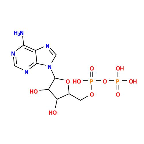 Mass: 427.201 Da / Num. of mol.: 5 / Source method: obtained synthetically / Formula: C10H15N5O10P2 / Comment: ADP, energy-carrying molecule*YM Mass: 427.201 Da / Num. of mol.: 5 / Source method: obtained synthetically / Formula: C10H15N5O10P2 / Comment: ADP, energy-carrying molecule*YM#5: Chemical | ChemComp-MG /   Mass: 24.305 Da / Num. of mol.: 5 / Source method: obtained synthetically / Formula: Mg Mass: 24.305 Da / Num. of mol.: 5 / Source method: obtained synthetically / Formula: MgHas ligand of interest | N | Has protein modification | N | |
|---|
-Experimental details
-Experiment
| Experiment | Method: ELECTRON MICROSCOPY |
|---|---|
| EM experiment | Aggregation state: FILAMENT / 3D reconstruction method: single particle reconstruction |
- Sample preparation
| Component | Name: Two AIP1 bound to the Barbed End of cofilin actin / Type: COMPLEX / Entity ID: #1-#3 / Source: MULTIPLE SOURCES |
|---|---|
| Molecular weight | Experimental value: NO |
| Source (natural) | Organism: Caenorhabditis elegans (invertebrata) |
| Source (recombinant) | Organism: Escherichia coli 'BL21-Gold(DE3)pLysS AG' (bacteria) |
| Buffer solution | pH: 7.5 |
| Specimen | Embedding applied: NO / Shadowing applied: NO / Staining applied: NO / Vitrification applied: YES |
| Vitrification | Instrument: FEI VITROBOT MARK IV / Cryogen name: ETHANE |
- Electron microscopy imaging
| Experimental equipment |  Model: Titan Krios / Image courtesy: FEI Company |
|---|---|
| Microscopy | Model: TFS KRIOS |
| Electron gun | Electron source:  FIELD EMISSION GUN / Accelerating voltage: 300 kV / Illumination mode: FLOOD BEAM FIELD EMISSION GUN / Accelerating voltage: 300 kV / Illumination mode: FLOOD BEAM |
| Electron lens | Mode: BRIGHT FIELD / Nominal magnification: 165000 X / Nominal defocus max: 2700 nm / Nominal defocus min: 500 nm |
| Image recording | Electron dose: 51.71 e/Å2 / Film or detector model: TFS FALCON 4i (4k x 4k) |
- Processing
| EM software |
| ||||||||||||||||||||||||
|---|---|---|---|---|---|---|---|---|---|---|---|---|---|---|---|---|---|---|---|---|---|---|---|---|---|
| CTF correction | Type: PHASE FLIPPING AND AMPLITUDE CORRECTION | ||||||||||||||||||||||||
| 3D reconstruction | Resolution: 2.69 Å / Resolution method: FSC 0.143 CUT-OFF / Num. of particles: 42984 / Symmetry type: POINT | ||||||||||||||||||||||||
| Refinement | Highest resolution: 2.69 Å Stereochemistry target values: REAL-SPACE (WEIGHTED MAP SUM AT ATOM CENTERS) | ||||||||||||||||||||||||
| Refine LS restraints |
|
