 Movie
Movie Controller


+ Open data

- Basic information
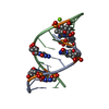
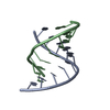
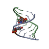
| Entry | Database: PDB / ID: 383d | ||||||||||||||||||
|---|---|---|---|---|---|---|---|---|---|---|---|---|---|---|---|---|---|---|---|
| Title | Hydration and recognition of methylated CPG steps in DNA | ||||||||||||||||||
 Components Components | DNA (5'-D(* Keywords KeywordsDNA / A-DNA / DOUBLE HELIX / MODIFIED DEOXYRIBONUCLEIC ACID | Function / homology | : / DNA |  Function and homology information Function and homology informationMethod |  X-RAY DIFFRACTION / SYNCHROTRON / MOLECULAR REPLACEMENT / Resolution: 1.7 Å X-RAY DIFFRACTION / SYNCHROTRON / MOLECULAR REPLACEMENT / Resolution: 1.7 Å  Authors AuthorsMayer-Jung, C. / Moras, D. / Timsit, Y. |  CitationJournal: Embo J. / Year: 1998 CitationJournal: Embo J. / Year: 1998Title: Hydration and Recognition of Methylated Cpg Steps in DNA Authors: Mayer-Jung, C. / Moras, D. / Timsit, Y. History |
|
- Structure visualization
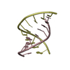
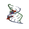
| Structure viewer | Molecule: MolmilJmol/JSmol |
|---|
- Downloads & links
-Download
| PDBx/mmCIF format | 383d.cif.gz | 21.9 KB | Display | PDBx/mmCIF format |
|---|---|---|---|---|
| PDB format | pdb383d.ent.gz | 15 KB | Display | PDB format |
| PDBx/mmJSON format | 383d.json.gz | Tree view | PDBx/mmJSON format | |
| Others |  Other downloads Other downloads |
-Validation report
| Arichive directory | https://data.pdbj.org/pub/pdb/validation_reports/83/383dftp://data.pdbj.org/pub/pdb/validation_reports/83/383d | HTTPS FTP |
|---|
-Related structure data


-Links
 PDBj
PDBj

- Assembly
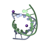
| Deposited unit | 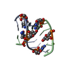
| ||||||||||
|---|---|---|---|---|---|---|---|---|---|---|---|
| 1 |
| ||||||||||
| Unit cell |
|
-Components
| #1: DNA chain | Mass: 3089.062 Da / Num. of mol.: 2 Source method: isolated from a genetically manipulated source References: NDB: ADJB110 #2: Chemical | ChemComp-MG / |   Mass: 24.305 Da / Num. of mol.: 1 / Source method: obtained synthetically / Formula: Mg Mass: 24.305 Da / Num. of mol.: 1 / Source method: obtained synthetically / Formula: Mg#3: Water | ChemComp-HOH / |  Mass: 18.015 Da / Num. of mol.: 87 / Source method: isolated from a natural source / Formula: H2O Mass: 18.015 Da / Num. of mol.: 87 / Source method: isolated from a natural source / Formula: H2O |
|---|
-Experimental details
-Experiment
| Experiment | Method: X-RAY DIFFRACTION / Number of used crystals: 1 |
|---|
- Sample preparation
| Crystal | Density Matthews: 1.98 Å3/Da / Density % sol: 37.85 % | ||||||||||||||||||||||||||||||
|---|---|---|---|---|---|---|---|---|---|---|---|---|---|---|---|---|---|---|---|---|---|---|---|---|---|---|---|---|---|---|---|
| Crystal grow | pH: 6 / Details: MPD, SPERMINE, MGCL2, pH 6.00 | ||||||||||||||||||||||||||||||
| Components of the solutions |
| ||||||||||||||||||||||||||||||
| Crystal grow | *PLUS Temperature: 20 ℃ / pH: 6 / Method: vapor diffusion, hanging drop | ||||||||||||||||||||||||||||||
| Components of the solutions | *PLUS
|
-Data collection
| Diffraction | Mean temperature: 270 K |
|---|---|
| Diffraction source | Source: SYNCHROTRON / Site: LURE  / Beamline: DW32 / Beamline: DW32 |
| Detector | Type: MARRESEARCH / Detector: IMAGE PLATE / Date: Mar 1, 1995 / Details: DOUBLE MIRRORS |
| Radiation | Monochromator: MIRROR + CHANNEL 4 / Protocol: SINGLE WAVELENGTH / Monochromatic (M) / Laue (L): M / Scattering type: x-ray |
| Radiation wavelength | Relative weight: 1 |
| Reflection | Resolution: 1.7→20 Å / Num. obs: 5804 / % possible obs: 94.4 % / Observed criterion σ(I): 1 / Redundancy: 4.6 % / Rmerge(I) obs: 0.038 |
| Reflection | *PLUS Highest resolution: 1.7 Å / Lowest resolution: 20 Å / % possible obs: 94.4 % / Observed criterion σ(I): 1 / Redundancy: 4.6 % |
- Processing
| Software |
| ||||||||||||||||||||||||||||||||||||||||||||||||||||||||||||
|---|---|---|---|---|---|---|---|---|---|---|---|---|---|---|---|---|---|---|---|---|---|---|---|---|---|---|---|---|---|---|---|---|---|---|---|---|---|---|---|---|---|---|---|---|---|---|---|---|---|---|---|---|---|---|---|---|---|---|---|---|---|
| Refinement | Method to determine structure: MOLECULAR REPLACEMENT Starting model: ISOMORPHOUS A-DNA Resolution: 1.7→7 Å / σ(F): 2
| ||||||||||||||||||||||||||||||||||||||||||||||||||||||||||||
| Refinement step | Cycle: LAST / Resolution: 1.7→7 Å
| ||||||||||||||||||||||||||||||||||||||||||||||||||||||||||||
| Refine LS restraints |
| ||||||||||||||||||||||||||||||||||||||||||||||||||||||||||||
| Software | *PLUS Name: X-PLOR / Version: 3.851 / Classification: refinement | ||||||||||||||||||||||||||||||||||||||||||||||||||||||||||||
| Refinement | *PLUS Highest resolution: 1.7 Å / Lowest resolution: 7 Å | ||||||||||||||||||||||||||||||||||||||||||||||||||||||||||||
| Solvent computation | *PLUS | ||||||||||||||||||||||||||||||||||||||||||||||||||||||||||||
| Displacement parameters | *PLUS | ||||||||||||||||||||||||||||||||||||||||||||||||||||||||||||
| Refine LS restraints | *PLUS
|
