 Movie
Movie Controller


+ Open data

- Basic information
| Entry | Database: PDB / ID: 36az | |||||||||||||||||||||
|---|---|---|---|---|---|---|---|---|---|---|---|---|---|---|---|---|---|---|---|---|---|---|
| Title | Structure of BA.1-S-RBD/2130WT/2196-S93Y | |||||||||||||||||||||
 Components Components |
| |||||||||||||||||||||
 Keywords Keywords | VIRAL PROTEIN / SARS-CoV-2 | |||||||||||||||||||||
| Function / homology |  Function and homology information Function and homology informationsymbiont-mediated disruption of host tissue / Maturation of spike protein / host cell surface / Translation of Structural Proteins / Virion Assembly and Release / Lectin pathway of complement activation / host extracellular region / symbiont-mediated-mediated suppression of host tetherin activity / structural constituent of virion / Induction of Cell-Cell Fusion ...symbiont-mediated disruption of host tissue / Maturation of spike protein / host cell surface / Translation of Structural Proteins / Virion Assembly and Release / Lectin pathway of complement activation / host extracellular region / symbiont-mediated-mediated suppression of host tetherin activity / structural constituent of virion / Induction of Cell-Cell Fusion / positive regulation of viral entry into host cell / Initial triggering of complement / membrane fusion / host cell endoplasmic reticulum-Golgi intermediate compartment membrane / Attachment and Entry / entry receptor-mediated virion attachment to host cell / receptor-mediated virion attachment to host cell / host cell surface receptor binding / symbiont-mediated suppression of host innate immune response / endocytosis involved in viral entry into host cell / receptor ligand activity / fusion of virus membrane with host plasma membrane / fusion of virus membrane with host endosome membrane / viral envelope / symbiont entry into host cell / virion attachment to host cell / host cell plasma membrane / SARS-CoV-2 activates/modulates innate and adaptive immune responses / virion membrane / membrane / identical protein binding / plasma membrane Similarity search - Function | |||||||||||||||||||||
| Biological species |  Homo sapiens (human) Homo sapiens (human) 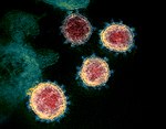 Severe acute respiratory syndrome coronavirus 2 Severe acute respiratory syndrome coronavirus 2 | |||||||||||||||||||||
| Method | ELECTRON MICROSCOPY / single particle reconstruction / cryo EM / Resolution: 3.9 Å | |||||||||||||||||||||
 Authors Authors | Du, J. / Pallesen, J. | |||||||||||||||||||||
| Funding support |  United States, 2items United States, 2items
| |||||||||||||||||||||
 Citation Citation | Journal: Res Sq / Year: 2026 Title: Structure-Guided Design of Therapeutic Antibodies Targeting SARS-CoV-2 Omicron Variants. Authors: Jesper Pallesen / Jianqiu Du / Yuanhan Wu / Sukanya Ghosh / Kelly Bayruns / Roopak Sadeesh / David Weiner Abstract: The ongoing evolution of SARS-CoV-2, particularly the emergence of Omicron subvariants, compromised the effectiveness of many therapeutic antibodies. In this study, we employed a structure-guided ...The ongoing evolution of SARS-CoV-2, particularly the emergence of Omicron subvariants, compromised the effectiveness of many therapeutic antibodies. In this study, we employed a structure-guided computational design strategy to systematically optimize the COV2-2196 antibody for improved neutralization of Omicron variants. Through iterative rounds of computational design and experimental validation, we identified key paratope mutations that restored and enhanced antibody binding and neutralization potency against resistant viral strains. Cryo-EM structural analysis revealed the molecular basis for these improvements, highlighting how targeted modifications can accommodate epitope changes introduced by viral evolution. Our approach demonstrates that effective antibody optimization can be achieved using accessible computational resources, providing a practical framework for rapid therapeutic development. These findings underscore the potential of structure-based design to address challenges posed by viral antigenic drift and support the development of broadly effective antibody therapeutics for emerging infectious diseases. | |||||||||||||||||||||
| History |
|
- Structure visualization
| Structure viewer | Molecule: MolmilJmol/JSmol |
|---|
- Downloads & links
-Download
| PDBx/mmCIF format | 36az.cif.gz | 164.6 KB | Display | PDBx/mmCIF format |
|---|---|---|---|---|
| PDB format | pdb36az.ent.gz | 115.2 KB | Display | PDB format |
| PDBx/mmJSON format | 36az.json.gz | Tree view | PDBx/mmJSON format | |
| Others |  Other downloads Other downloads |
-Validation report
| Arichive directory | https://data.pdbj.org/pub/pdb/validation_reports/6a/36azftp://data.pdbj.org/pub/pdb/validation_reports/6a/36az | HTTPS FTP |
|---|
-Related structure data
| Related structure data |  77344MC  36bbC M: map data used to model this data C: citing same article ( |
|---|---|
| Similar structure data |


-Links
 PDBj
PDBj
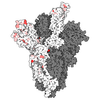
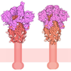

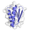

- Assembly
| Deposited unit | 
|
|---|---|
| 1 |
|
-Components
-Antibody , 4 types, 4 molecules HLBC
| #1: Antibody | Mass: 52514.156 Da / Num. of mol.: 1 Source method: isolated from a genetically manipulated source Source: (gene. exp.) Homo sapiens (human) / Production host: Homo sapiens (human) |
|---|---|
| #2: Antibody | Mass: 26326.418 Da / Num. of mol.: 1 Source method: isolated from a genetically manipulated source Source: (gene. exp.) Homo sapiens (human) / Production host: Homo sapiens (human) |
| #4: Antibody | Mass: 25898.893 Da / Num. of mol.: 1 Source method: isolated from a genetically manipulated source Source: (gene. exp.) Homo sapiens (human) / Production host: Homo sapiens (human) |
| #5: Antibody | Mass: 51845.508 Da / Num. of mol.: 1 Source method: isolated from a genetically manipulated source Source: (gene. exp.) Homo sapiens (human) / Production host: Homo sapiens (human) |
-Protein / Sugars , 2 types, 2 molecules A
| #3: Protein | Mass: 25145.273 Da / Num. of mol.: 1 / Fragment: receptor binding domain Source method: isolated from a genetically manipulated source Source: (gene. exp.) Severe acute respiratory syndrome coronavirus 2Strain: Omicron BA.1 / Gene: S, 2 / Production host: Homo sapiens (human) / References: UniProt: P0DTC2 |
|---|---|
| #6: Sugar | ChemComp-NAG /  Type: D-saccharide, beta linking / Mass: 221.208 Da / Num. of mol.: 1 / Source method: obtained synthetically / Formula: C8H15NO6 Type: D-saccharide, beta linking / Mass: 221.208 Da / Num. of mol.: 1 / Source method: obtained synthetically / Formula: C8H15NO6 |
-Details
| Has ligand of interest | N |
|---|---|
| Has protein modification | Y |
-Experimental details
-Experiment
| Experiment | Method: ELECTRON MICROSCOPY |
|---|---|
| EM experiment | Aggregation state: PARTICLE / 3D reconstruction method: single particle reconstruction |
- Sample preparation
| Component |
| ||||||||||||||||||||||||||||||
|---|---|---|---|---|---|---|---|---|---|---|---|---|---|---|---|---|---|---|---|---|---|---|---|---|---|---|---|---|---|---|---|
| Source (natural) |
| ||||||||||||||||||||||||||||||
| Source (recombinant) |
| ||||||||||||||||||||||||||||||
| Buffer solution | pH: 7.4 | ||||||||||||||||||||||||||||||
| Specimen | Embedding applied: NO / Shadowing applied: NO / Staining applied: NO / Vitrification applied: YES | ||||||||||||||||||||||||||||||
| Vitrification | Cryogen name: ETHANE |
- Electron microscopy imaging
| Experimental equipment |  Model: Titan Krios / Image courtesy: FEI Company |
|---|---|
| Microscopy | Model: TFS KRIOS |
| Electron gun | Electron source:  FIELD EMISSION GUN / Accelerating voltage: 300 kV / Illumination mode: SPOT SCAN FIELD EMISSION GUN / Accelerating voltage: 300 kV / Illumination mode: SPOT SCAN |
| Electron lens | Mode: BRIGHT FIELD / Nominal defocus max: 2500 nm / Nominal defocus min: 500 nm |
| Image recording | Electron dose: 58.6 e/Å2 / Film or detector model: GATAN K3 (6k x 4k) |
- Processing
| EM software |
| |||||||||
|---|---|---|---|---|---|---|---|---|---|---|
| CTF correction | Type: NONE | |||||||||
| 3D reconstruction | Resolution: 3.9 Å / Resolution method: FSC 0.143 CUT-OFF / Num. of particles: 243248 / Symmetry type: POINT |
