Movie
 Controller


[English] 日本語
 Yorodumi
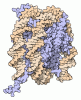
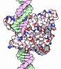
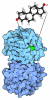
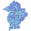
- PDB-36ap: Mycobacterium tuberculosis DNA Gyrase catalytic core in complex w... -
+ Open data

- Basic information
| Entry | Database: PDB / ID: 36ap | |||||||||
|---|---|---|---|---|---|---|---|---|---|---|
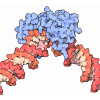
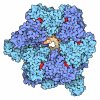
| Title | Mycobacterium tuberculosis DNA Gyrase catalytic core in complex with DNA and indole-3-acetyl-L-aspartic acid | |||||||||
 Components |
| |||||||||
 Keywords | OXIDOREDUCTASE / Mycobacterium tuberculosis / InhA / Inhibitory adduct / Resistance / Mutation | |||||||||
| Function / homology |  Function and homology information DNA negative supercoiling activity / DNA topoisomerase type II (double strand cut, ATP-hydrolyzing) complex / DNA topoisomerase type II (double strand cut, ATP-hydrolyzing) activity / DNA topoisomerase (ATP-hydrolysing) / DNA topological change / DNA-templated DNA replication / chromosome / magnesium ion binding / ATP hydrolysis activity / DNA binding ...DNA negative supercoiling activity / DNA topoisomerase type II (double strand cut, ATP-hydrolyzing) complex / DNA topoisomerase type II (double strand cut, ATP-hydrolyzing) activity / DNA topoisomerase (ATP-hydrolysing) / DNA topological change / DNA-templated DNA replication / chromosome / magnesium ion binding / ATP hydrolysis activity / DNA binding / ATP binding / plasma membrane / cytoplasm Similarity search - Function | |||||||||
| Biological species |  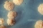 Mycobacterium tuberculosis (bacteria) synthetic construct (others) | |||||||||
| Method | ELECTRON MICROSCOPY / single particle reconstruction / cryo EM / Resolution: 2.64 Å | |||||||||
 Authors | Rossini, N.O. / Dias, M.V.B. / Coureux, P.D. | |||||||||
| Funding support |  Brazil, 2items
| |||||||||
 Citation | Journal: Eur J Med Chem / Year: 2026 Title: Fragment-based drug discovery strategy reveals an unprecedented allosteric binding site in Mycobacterium tuberculosis DNA gyrase. Authors: Sabrina T Shiroma / Nicolas O Rossini / Jillian F Armenia / Catharina S Silva / Paulo C T Souza / Pierre-Damien Coureux / Neil Osheroff / Marcio V B Dias /   Abstract: Tuberculosis remains a serious public health threat and the leading infectious cause of death worldwide. Tuberculosis treatment requires at least four antimicrobial drugs and is associated with ...Tuberculosis remains a serious public health threat and the leading infectious cause of death worldwide. Tuberculosis treatment requires at least four antimicrobial drugs and is associated with adverse effects and prolonged therapy, compromising adherence and increasing the risk of drug resistance. Therefore, developing new antitubercular therapies remains essential. Mycobacterium tuberculosis, the primary etiological agent of tuberculosis, lacks topoisomerase IV, making DNA gyrase its sole type II topoisomerase and an attractive drug target. Although fluoroquinolones such as moxifloxacin are used to treat tuberculosis, resistance has limited their effectiveness. Here, we applied a fragment-based drug discovery approach to identify and validate low-affinity compounds targeting M. tuberculosis DNA gyrase as starting points for inhibitor development. Using complementary biophysical and biochemical techniques, we identified five compounds that bind DNA gyrase and inhibit DNA supercoiling and DNA cleavage activities. These compounds interfere with early steps of the gyrase catalytic cycle, likely by blocking conformational changes required for DNA cleavage. The cryo-electron microscopy structure of DNA gyrase in complex with the most potent compound revealed binding near the GyrA-GyrB interface and extensive conformational changes in the GyrB TOPRIM domain. These findings provide a foundation for developing antitubercular agents with a novel mechanism of action. | |||||||||
| History |
|
- Structure visualization
| Structure viewer | Molecule: MolmilJmol/JSmol |
|---|
- Downloads & links
-Download
| PDBx/mmCIF format | 36ap.cif.gz | 317.6 KB | Display | PDBx/mmCIF format |
|---|---|---|---|---|
| PDB format | pdb36ap.ent.gz | 251.8 KB | Display | PDB format |
| PDBx/mmJSON format | 36ap.json.gz | Tree view | PDBx/mmJSON format | |
| Others |  Other downloads |
-Validation report
| Arichive directory | https://data.pdbj.org/pub/pdb/validation_reports/6a/36apftp://data.pdbj.org/pub/pdb/validation_reports/6a/36ap | HTTPS FTP |
|---|
-Related structure data
| Related structure data |  77331MC M: map data used to model this data C: citing same article ( |
|---|---|
| Similar structure data |
-Links
 PDBj
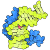
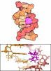
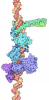
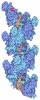
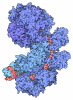
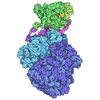
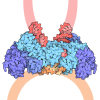
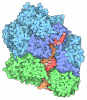
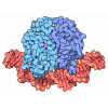
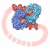
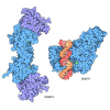
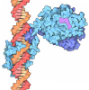

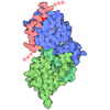
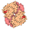
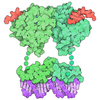
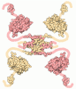
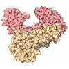
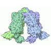
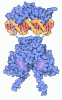
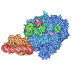
- Assembly
| Deposited unit | 
|
|---|---|
| 1 |
|
-Components
-DNA gyrase subunit ... , 2 types, 4 molecules ACBD
| #1: Protein | Mass: 55928.262 Da / Num. of mol.: 2 Source method: isolated from a genetically manipulated source Source: (gene. exp.) Mycobacterium tuberculosis (bacteria) / Gene: gyrA, Rv0006, MTCY10H4.04 / Production host:  Escherichia coli (E. coli) References: UniProt: P9WG47, DNA topoisomerase (ATP-hydrolysing) #2: Protein | Mass: 28185.334 Da / Num. of mol.: 2 Source method: isolated from a genetically manipulated source Source: (gene. exp.) Mycobacterium tuberculosis (bacteria) / Gene: gyrB, Rv0005, MTCY10H4.03 / Production host: Escherichia coli (E. coli)References: UniProt: P9WG45, DNA topoisomerase (ATP-hydrolysing) |
|---|
-DNA chain , 4 types, 4 molecules EHFG
| #3: DNA chain | Mass: 3357.223 Da / Num. of mol.: 1 / Source method: obtained synthetically / Source: (synth.) synthetic construct (others) |
|---|---|
| #4: DNA chain | Mass: 3044.017 Da / Num. of mol.: 1 / Source method: obtained synthetically / Source: (synth.) synthetic construct (others) |
| #5: DNA chain | Mass: 2401.604 Da / Num. of mol.: 1 / Source method: obtained synthetically / Source: (synth.) synthetic construct (others) |
| #6: DNA chain | Mass: 2779.848 Da / Num. of mol.: 1 / Source method: obtained synthetically / Source: (synth.) synthetic construct (others) |
-Non-polymers , 2 types, 6 molecules 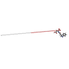

| #7: Chemical | ChemComp-IAD /  Mass: 290.271 Da / Num. of mol.: 4 / Source method: obtained synthetically / Formula: C14H14N2O5 / Feature type: SUBJECT OF INVESTIGATION #8: Chemical |  Mass: 24.305 Da / Num. of mol.: 2 / Source method: obtained synthetically / Formula: Mg |
|---|
-Details
| Has ligand of interest | Y |
|---|---|
| Has protein modification | N |
-Experimental details
-Experiment
| Experiment | Method: ELECTRON MICROSCOPY |
|---|---|
| EM experiment | Aggregation state: PARTICLE / 3D reconstruction method: single particle reconstruction |
- Sample preparation
| Component | Name: Tetrameric structure of APO InhA mutant I95P / Type: COMPLEX / Entity ID: #1 / Source: RECOMBINANT | |||||||||||||||
|---|---|---|---|---|---|---|---|---|---|---|---|---|---|---|---|---|
| Molecular weight | Value: 0.114 MDa / Experimental value: YES | |||||||||||||||
| Source (natural) | Organism: Mycobacterium tuberculosis (bacteria) | |||||||||||||||
| Source (recombinant) | Organism: Escherichia coli (E. coli) | |||||||||||||||
| Buffer solution | pH: 7 | |||||||||||||||
| Buffer component |
| |||||||||||||||
| Specimen | Conc.: 0.1 mg/ml / Embedding applied: NO / Shadowing applied: NO / Staining applied: NO / Vitrification applied: YES | |||||||||||||||
| Vitrification | Instrument: FEI VITROBOT MARK IV / Cryogen name: ETHANE / Humidity: 95 % / Chamber temperature: 298 K |
- Electron microscopy imaging
| Experimental equipment | 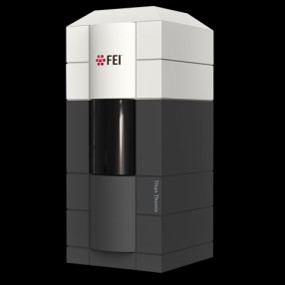 Model: Titan Krios / Image courtesy: FEI Company |
|---|---|
| Microscopy | Model: TFS KRIOS |
| Electron gun | Electron source:  FIELD EMISSION GUN / Accelerating voltage: 300 kV / Illumination mode: FLOOD BEAM |
| Electron lens | Mode: BRIGHT FIELD / Nominal magnification: 130000 X / Nominal defocus max: 2500 nm / Nominal defocus min: 800 nm / Cs: 2.7 mm |
| Specimen holder | Cryogen: NITROGEN |
| Image recording | Electron dose: 52 e/Å2 / Film or detector model: GATAN K3 (6k x 4k) |
| EM imaging optics | Energyfilter name: GIF Quantum LS |
- Processing
| EM software |
| ||||||||||||||||||||||||||||
|---|---|---|---|---|---|---|---|---|---|---|---|---|---|---|---|---|---|---|---|---|---|---|---|---|---|---|---|---|---|
| CTF correction | Type: PHASE FLIPPING AND AMPLITUDE CORRECTION | ||||||||||||||||||||||||||||
| Particle selection | Num. of particles selected: 13185442 | ||||||||||||||||||||||||||||
| 3D reconstruction | Resolution: 2.64 Å / Resolution method: FSC 0.143 CUT-OFF / Num. of particles: 61358 / Symmetry type: POINT | ||||||||||||||||||||||||||||
| Refinement | Highest resolution: 2.64 Å Stereochemistry target values: REAL-SPACE (WEIGHTED MAP SUM AT ATOM CENTERS) | ||||||||||||||||||||||||||||
| Refine LS restraints |
|