 Movie
Movie Controller

[English] 日本語
 Yorodumi
- PDB-31np: Crystal structure of the complex of galectin-8N-2,6-anhydro-3-deo... -
+ Open data

- Basic information
| Entry | Database: PDB / ID: 31np | ||||||
|---|---|---|---|---|---|---|---|
| Title | Crystal structure of the complex of galectin-8N-2,6-anhydro-3-deoxy-3-S-(beta-D-galactopyranosyl)-3-thio-D-glycero-L-altro-heptonamide | ||||||
 Components Components | Galectin-8 | ||||||
 Keywords Keywords | SUGAR BINDING PROTEIN / galectin-8N | ||||||
| Function / homology |  Function and homology information Function and homology informationlymphatic endothelial cell migration / xenophagy / cellular response to virus / integrin binding / carbohydrate binding / cytoplasmic vesicle / : / membrane / cytosol / cytoplasm Similarity search - Function | ||||||
| Biological species |  Homo sapiens (human) Homo sapiens (human) | ||||||
| Method |  X-RAY DIFFRACTION / SYNCHROTRON / MOLECULAR REPLACEMENT / Resolution: 1.78 Å X-RAY DIFFRACTION / SYNCHROTRON / MOLECULAR REPLACEMENT / Resolution: 1.78 Å | ||||||
 Authors Authors | Tsagkarakou, A.S. / Kantsadi, A.L. / Leonidas, D.D. | ||||||
| Funding support | 1items
| ||||||
 Citation Citation | Journal: Chemmedchem / Year: 2026 Title: Structural and Biophysical Characterization of C-Glycosylic 1,2-Thiodisaccharides Reveals Determinants of Selective Binding to Galectin-7 and Galectin-8N. Authors: Tsagkarakou, A.S. / Kantsadi, A.L. / Theodoridou, V.I. / Veliotis, N. / Lazar, L. / Jozsef, J. / Juhasz, L. / Kontopidis, G. / Leffler, H. / Nilsson, U.J. / Somsak, L. / Leonidas, D.D. | ||||||
| History |
|
- Structure visualization
| Structure viewer | Molecule: MolmilJmol/JSmol |
|---|
- Downloads & links
-Download
| PDBx/mmCIF format | 31np.cif.gz | 98.9 KB | Display | PDBx/mmCIF format |
|---|---|---|---|---|
| PDB format | pdb31np.ent.gz | Display | PDB format | |
| PDBx/mmJSON format | 31np.json.gz | Tree view | PDBx/mmJSON format | |
| Others |  Other downloads Other downloads |
-Validation report
| Arichive directory | https://data.pdbj.org/pub/pdb/validation_reports/1n/31npftp://data.pdbj.org/pub/pdb/validation_reports/1n/31np | HTTPS FTP |
|---|
-Related structure data
| Related structure data |  31nvC  31nwC  31ml C: citing same article ( |
|---|---|
| Similar structure data |
-Links
 PDBj
PDBj- Assembly
| Deposited unit | 
| ||||||||||||
|---|---|---|---|---|---|---|---|---|---|---|---|---|---|
| 1 |
| ||||||||||||
| Unit cell |
|
-Components
| #1: Protein | Mass: 35854.055 Da / Num. of mol.: 1 Source method: isolated from a genetically manipulated source Source: (gene. exp.) Homo sapiens (human) / Gene: LGALS8 / Production host:   Escherichia coli BL21(DE3) (bacteria) / References: UniProt: O00214 Escherichia coli BL21(DE3) (bacteria) / References: UniProt: O00214 | ||||||||||
|---|---|---|---|---|---|---|---|---|---|---|---|
| #2: Chemical | 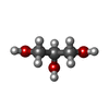 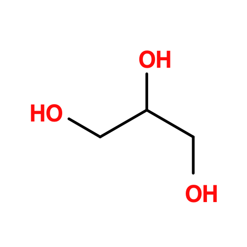 Mass: 92.094 Da / Num. of mol.: 2 / Source method: obtained synthetically / Formula: C3H8O3 Mass: 92.094 Da / Num. of mol.: 2 / Source method: obtained synthetically / Formula: C3H8O3#3: Sugar | ChemComp-YIO / | 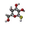 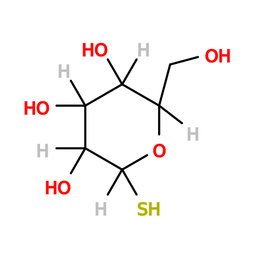 Type: D-saccharide, beta linking / Mass: 196.221 Da / Num. of mol.: 1 / Source method: obtained synthetically / Formula: C6H12O5S / Feature type: SUBJECT OF INVESTIGATION Type: D-saccharide, beta linking / Mass: 196.221 Da / Num. of mol.: 1 / Source method: obtained synthetically / Formula: C6H12O5S / Feature type: SUBJECT OF INVESTIGATION#4: Chemical | ChemComp-A1KB4 / ( | Mass: 207.181 Da / Num. of mol.: 1 / Source method: obtained synthetically / Formula: C7H13NO6 / Feature type: SUBJECT OF INVESTIGATION #5: Water | ChemComp-HOH / |  Mass: 18.015 Da / Num. of mol.: 80 / Source method: isolated from a natural source / Formula: H2O Mass: 18.015 Da / Num. of mol.: 80 / Source method: isolated from a natural source / Formula: H2OHas ligand of interest | Y | Has protein modification | N | |
-Experimental details
-Experiment
| Experiment | Method: X-RAY DIFFRACTION / Number of used crystals: 1 |
|---|
- Sample preparation
| Crystal grow | Temperature: 289 K / Method: vapor diffusion, hanging drop / pH: 5.6 Details: Protein at 10 mg/mL in PBS (pH 7.4) was mixed 1:1 with reservoir solution containing 0.1 M Tris/NaOAc (pH 5.6) 20% (v/v) 2-propanol and 20% PEG 4000. |
|---|
-Data collection
| Diffraction | Mean temperature: 100 K / Serial crystal experiment: N |
|---|---|
| Diffraction source | Source: SYNCHROTRON / Site: PETRA III, EMBL c/o DESY  / Beamline: P13 (MX1) / Wavelength: 0.976 Å / Beamline: P13 (MX1) / Wavelength: 0.976 Å |
| Detector | Type: DECTRIS PILATUS 6M / Detector: PIXEL / Date: Mar 14, 2022 |
| Radiation | Monochromator: Mirrors / Protocol: SINGLE WAVELENGTH / Monochromatic (M) / Laue (L): M / Scattering type: x-ray |
| Radiation wavelength | Wavelength: 0.976 Å / Relative weight: 1 |
| Reflection | Resolution: 1.78→62 Å / Num. obs: 14804 / % possible obs: 99 % / Observed criterion σ(F): 0 / Observed criterion σ(I): -3 / Redundancy: 6.8 % / Biso Wilson estimate: 32.97 Å2 / CC1/2: 0.998 / Rmerge(I) obs: 0.077 / Net I/σ(I): 11.5 |
| Reflection shell | Resolution: 1.78→1.82 Å / Redundancy: 6.8 % / Rmerge(I) obs: 0.803 / Mean I/σ(I) obs: 2.3 / Num. unique obs: 832 / CC1/2: 0.797 / % possible all: 99 |
- Processing
| Software |
| ||||||||||||||||||||||||||||||||||||||||||
|---|---|---|---|---|---|---|---|---|---|---|---|---|---|---|---|---|---|---|---|---|---|---|---|---|---|---|---|---|---|---|---|---|---|---|---|---|---|---|---|---|---|---|---|
| Refinement | Method to determine structure: MOLECULAR REPLACEMENT / Resolution: 1.78→27.84 Å / SU ML: 0.1896 / Cross valid method: FREE R-VALUE / σ(F): 1.35 / Phase error: 25.658 Stereochemistry target values: GeoStd + Monomer Library + CDL v1.2
| ||||||||||||||||||||||||||||||||||||||||||
| Solvent computation | Shrinkage radii: 0.9 Å / VDW probe radii: 1.1 Å / Solvent model: FLAT BULK SOLVENT MODEL | ||||||||||||||||||||||||||||||||||||||||||
| Displacement parameters | Biso mean: 40.92 Å2 | ||||||||||||||||||||||||||||||||||||||||||
| Refinement step | Cycle: LAST / Resolution: 1.78→27.84 Å
| ||||||||||||||||||||||||||||||||||||||||||
| Refine LS restraints |
| ||||||||||||||||||||||||||||||||||||||||||
| LS refinement shell |
| ||||||||||||||||||||||||||||||||||||||||||
| Refinement TLS params. | Method: refined / Origin x: 19.368071663094 Å / Origin y: 5.110254410653 Å / Origin z: 10.888141752524 Å
| ||||||||||||||||||||||||||||||||||||||||||
| Refinement TLS group | Selection details: all |
