Biotechnology and Biological Sciences Research Council (BBSRC)
BB/P02002X/1
United Kingdom
Citation
Journal: IUCrJ / Year: 2026 Title: How cryoEM has advanced our understanding of bacteriophages and bacteriocins targeting Clostridioides difficile. Authors: Per A Bullough / Jason S Wilson / Hannah L Berry / Robert P Fagan / Abstract: We review the structural and functional characteristics of bacteriophages and bacteriocins (diffocins) that specifically target Clostridioides difficile, a significant healthcare concern due to its ...We review the structural and functional characteristics of bacteriophages and bacteriocins (diffocins) that specifically target Clostridioides difficile, a significant healthcare concern due to its role in nosocomial infections. The advent of modern cryogenic electron microscopy (cryoEM) has revolutionized our understanding of these contractile injection systems, providing high-resolution insights into their mechanisms. We compare the structures of C. difficile phages and diffocins, highlighting their adaptations for penetrating the Gram-positive bacterial cell envelope - including the cell membrane, cell wall and proteinaceous surface layer. Diffocins, simpler in structure, utilize a combination of mechanical and enzymatic strategies, while some phages like ΦCD508 may rely on mechanical force alone. This review delves into the assembly and function of key components such as the contractile sheath, baseplate and receptor-binding proteins, offering a framework for engineering precision antimicrobials. We also present new experimental results, including refined cryoEM structures of the ΦCD508 pre- and post-contracted tail, a novel spontaneously contracted conformation and an X-ray crystal structure of a phage receptor-binding protein domain. This work underscores the potential of cryoEM in advancing our understanding of phage biology and its applications in developing targeted therapies against C. difficile.
Method to determine structure: SAD / Resolution: 1.42→46.789 Å / Cor.coef. Fo:Fc: 0.97 / Cor.coef. Fo:Fc free: 0.965 / SU B: 1.198 / SU ML: 0.043 / Cross valid method: FREE R-VALUE / ESU R: 0.049 / ESU R Free: 0.05 Details: Hydrogens have been added in their riding positions
Rfactor
Num. reflection
% reflection
Rfree
0.2017
5081
4.923 %
Rwork
0.1852
98125
-
all
0.186
-
-
obs
-
103206
99.788 %
Solvent computation
Ion probe radii: 0.8 Å / Shrinkage radii: 0.8 Å / VDW probe radii: 1.2 Å / Solvent model: MASK BULK SOLVENT
Displacement parameters
Biso mean: 20.608 Å2
Baniso -1
Baniso -2
Baniso -3
1-
0.183 Å2
0 Å2
-0.91 Å2
2-
-
-0.264 Å2
0 Å2
3-
-
-
0.372 Å2
Refinement step
Cycle: LAST / Resolution: 1.42→46.789 Å
Protein
Nucleic acid
Ligand
Solvent
Total
Num. atoms
2579
0
5
239
2823
Refine LS restraints
Refine-ID
Type
Dev ideal
Dev ideal target
Number
X-RAY DIFFRACTION
r_bond_refined_d
0.014
0.012
2704
X-RAY DIFFRACTION
r_bond_other_d
0.001
0.017
2321
X-RAY DIFFRACTION
r_angle_refined_deg
1.881
1.642
3687
X-RAY DIFFRACTION
r_angle_other_deg
1.565
1.575
5405
X-RAY DIFFRACTION
r_dihedral_angle_1_deg
7.393
5
334
X-RAY DIFFRACTION
r_dihedral_angle_2_deg
34.15
24.437
142
X-RAY DIFFRACTION
r_dihedral_angle_3_deg
11.546
15
415
X-RAY DIFFRACTION
r_dihedral_angle_4_deg
6.674
15
4
X-RAY DIFFRACTION
r_chiral_restr
0.101
0.2
355
X-RAY DIFFRACTION
r_gen_planes_refined
0.011
0.02
3078
X-RAY DIFFRACTION
r_gen_planes_other
0.001
0.02
618
X-RAY DIFFRACTION
r_nbd_refined
0.215
0.2
409
X-RAY DIFFRACTION
r_symmetry_nbd_other
0.189
0.2
2015
X-RAY DIFFRACTION
r_nbtor_refined
0.186
0.2
1331
X-RAY DIFFRACTION
r_symmetry_nbtor_other
0.084
0.2
1297
X-RAY DIFFRACTION
r_xyhbond_nbd_refined
0.162
0.2
136
X-RAY DIFFRACTION
r_metal_ion_refined
0.111
0.2
4
X-RAY DIFFRACTION
r_symmetry_nbd_refined
0.166
0.2
6
X-RAY DIFFRACTION
r_nbd_other
0.173
0.2
27
X-RAY DIFFRACTION
r_symmetry_xyhbond_nbd_refined
0.091
0.2
15
X-RAY DIFFRACTION
r_mcbond_it
2.023
1.985
1294
X-RAY DIFFRACTION
r_mcbond_other
2.021
1.983
1293
X-RAY DIFFRACTION
r_mcangle_it
2.793
2.966
1618
X-RAY DIFFRACTION
r_mcangle_other
2.793
2.968
1619
X-RAY DIFFRACTION
r_scbond_it
3.839
2.316
1409
X-RAY DIFFRACTION
r_scbond_other
3.842
2.318
1410
X-RAY DIFFRACTION
r_scangle_it
5.59
3.344
2061
X-RAY DIFFRACTION
r_scangle_other
5.589
3.345
2062
X-RAY DIFFRACTION
r_lrange_it
7.145
23.64
2993
X-RAY DIFFRACTION
r_lrange_other
7.127
23.398
2964
LS refinement shell
Refine-ID: X-RAY DIFFRACTION / Total num. of bins used: 20
Resolution (Å)
Rfactor Rfree
Num. reflection Rfree
Rfactor Rwork
Num. reflection Rwork
Rfactor all
Num. reflection all
Fsc free
Fsc work
% reflection obs (%)
WRfactor Rwork
1.42-1.457
0.329
382
0.333
7225
0.332
7608
0.436
0.432
99.9869
0.329
1.457-1.497
0.339
374
0.332
7051
0.332
7425
0.652
0.648
100
0.322
1.497-1.54
0.298
354
0.305
6873
0.305
7231
0.766
0.775
99.9447
0.287
1.54-1.587
0.305
333
0.284
6649
0.285
6985
0.814
0.822
99.957
0.259
1.587-1.639
0.289
321
0.265
6485
0.266
6811
0.848
0.855
99.9266
0.234
1.639-1.697
0.263
335
0.24
6261
0.241
6599
0.889
0.891
99.9545
0.209
1.697-1.761
0.242
320
0.219
5991
0.22
6317
0.902
0.912
99.905
0.189
1.761-1.833
0.224
292
0.189
5802
0.191
6094
0.928
0.935
100
0.169
1.833-1.914
0.191
299
0.172
5601
0.173
5902
0.948
0.955
99.9661
0.157
1.914-2.007
0.184
270
0.168
5334
0.169
5604
0.959
0.962
100
0.157
2.007-2.116
0.182
280
0.166
5038
0.167
5322
0.959
0.965
99.9248
0.159
2.116-2.244
0.189
227
0.167
4857
0.168
5099
0.954
0.961
99.7058
0.164
2.244-2.398
0.193
264
0.166
4459
0.167
4727
0.955
0.96
99.9154
0.168
2.398-2.59
0.188
215
0.167
4228
0.168
4453
0.96
0.962
99.7754
0.172
2.59-2.836
0.177
176
0.17
3871
0.17
4070
0.955
0.958
99.4349
0.182
2.836-3.169
0.195
190
0.176
3468
0.177
3691
0.946
0.954
99.1059
0.197
3.169-3.657
0.181
145
0.166
3115
0.166
3288
0.959
0.964
99.1484
0.19
3.657-4.472
0.155
136
0.152
2613
0.152
2780
0.973
0.973
98.8849
0.188
4.472-6.296
0.193
110
0.165
2049
0.166
2169
0.973
0.975
99.539
0.211
6.296-46.789
0.233
58
0.242
1154
0.242
1246
0.952
0.94
97.2713
0.301
+
About Yorodumi
-
News
-
Feb 9, 2022. New format data for meta-information of EMDB entries
New format data for meta-information of EMDB entries
Version 3 of the EMDB header file is now the official format.
The previous official version 1.9 will be removed from the archive.
In the structure databanks used in Yorodumi, some data are registered as the other names, "COVID-19 virus" and "2019-nCoV". Here are the details of the virus and the list of structure data.
Jan 31, 2019. EMDB accession codes are about to change! (news from PDBe EMDB page)
EMDB accession codes are about to change! (news from PDBe EMDB page)
The allocation of 4 digits for EMDB accession codes will soon come to an end. Whilst these codes will remain in use, new EMDB accession codes will include an additional digit and will expand incrementally as the available range of codes is exhausted. The current 4-digit format prefixed with “EMD-” (i.e. EMD-XXXX) will advance to a 5-digit format (i.e. EMD-XXXXX), and so on. It is currently estimated that the 4-digit codes will be depleted around Spring 2019, at which point the 5-digit format will come into force.
The EM Navigator/Yorodumi systems omit the EMD- prefix.
Related info.:Q: What is EMD? / ID/Accession-code notation in Yorodumi/EM Navigator
Yorodumi is a browser for structure data from EMDB, PDB, SASBDB, etc.
This page is also the successor to EM Navigator detail page, and also detail information page/front-end page for Omokage search.
The word "yorodu" (or yorozu) is an old Japanese word meaning "ten thousand". "mi" (miru) is to see.
Related info.:EMDB / PDB / SASBDB / Comparison of 3 databanks / Yorodumi Search / Aug 31, 2016. New EM Navigator & Yorodumi / Yorodumi Papers / Jmol/JSmol / Function and homology information / Changes in new EM Navigator and Yorodumi
 Movie
Movie Controller


 Open data
 Basic information
 Components
 Keywords
 Function and homology information
 Clostridioides difficile R20291 (bacteria)
 X-RAY DIFFRACTION /
  Authors
 United Kingdom, 1items
  Citation
 Structure visualization
 Downloads & links
 Other downloads


 PDBj
PDBj Assembly
 Escherichia coli BL21 (bacteria) / References: UniProt: A0AB74R428
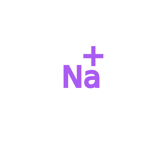 Mass: 22.990 Da / Num. of mol.: 1 / Source method: obtained synthetically / Formula: Na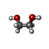
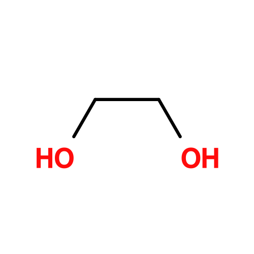 Mass: 62.068 Da / Num. of mol.: 1 / Source method: obtained synthetically / Formula: C2H6O2
 Mass: 18.015 Da / Num. of mol.: 239 / Source method: isolated from a natural source / Formula: H2O
 Sample preparation
 Processing