 Movie
Movie Controller

[English] 日本語
 Yorodumi
- PDB-31gl: Prescottella amidase, S181A mutant, diethyl toluene-2,4-dicarbama... -
+ Open data

- Basic information
| Entry | Database: PDB / ID: 31gl | ||||||
|---|---|---|---|---|---|---|---|
| Title | Prescottella amidase, S181A mutant, diethyl toluene-2,4-dicarbamate soak | ||||||
 Components Components | amidase | ||||||
 Keywords Keywords | HYDROLASE / Urethanase | ||||||
| Function / homology |  Function and homology information Function and homology information | ||||||
| Biological species |  Prescottella equi (bacteria) Prescottella equi (bacteria) | ||||||
| Method |  X-RAY DIFFRACTION / SYNCHROTRON / MOLECULAR REPLACEMENT / Resolution: 1.2 Å X-RAY DIFFRACTION / SYNCHROTRON / MOLECULAR REPLACEMENT / Resolution: 1.2 Å | ||||||
 Authors Authors | Bloch, Y. / Panneerselvam, S. | ||||||
| Funding support | European Union, 1items
| ||||||
 Citation Citation | Journal: To Be Published Title: Crystallographic exploration of a Prescottella sp. amidase as a model system for urethanase activity. Authors: Bloch, Y. / Panneerselvam, S. / Schneider, T.R. #1: Journal: Appl Microbiol Biotechnol / Year: 2006 Title: Isolation of a bacterium that degrades urethane compounds and characterization of its urethane hydrolase. Authors: Akutsu-Shigeno, Y. / Adachi, Y. / Yamada, C. / Toyoshima, K. / Nomura, N. / Uchiyama, H. / Nakajima-Kambe, T. | ||||||
| History |
|
- Structure visualization
| Structure viewer | Molecule: MolmilJmol/JSmol |
|---|
- Downloads & links
-Download
| PDBx/mmCIF format | 31gl.cif.gz | 585.8 KB | Display | PDBx/mmCIF format |
|---|---|---|---|---|
| PDB format | pdb31gl.ent.gz | Display | PDB format | |
| PDBx/mmJSON format | 31gl.json.gz | Tree view | PDBx/mmJSON format | |
| Others |  Other downloads Other downloads |
-Validation report
| Arichive directory | https://data.pdbj.org/pub/pdb/validation_reports/1g/31glftp://data.pdbj.org/pub/pdb/validation_reports/1g/31gl | HTTPS FTP |
|---|
-Related structure data


-Links
 PDBj
PDBj- Assembly
| Deposited unit | 
| ||||||||
|---|---|---|---|---|---|---|---|---|---|
| 1 | 
| ||||||||
| 2 | 
| ||||||||
| Unit cell |
|
-Components
-Protein , 1 types, 2 molecules AB
| #1: Protein | Mass: 50901.906 Da / Num. of mol.: 2 / Mutation: S181A Source method: isolated from a genetically manipulated source Details: residues part of cloning and purification strategy residues His6tag residues + HRV 3C tag residues removed by proteolytic digest actual protein starts from residue 21 which is natively ...Details: residues part of cloning and purification strategy residues His6tag residues + HRV 3C tag residues removed by proteolytic digest actual protein starts from residue 21 which is natively residue 2 S181A mismatch is mutation in catalytic residue Source: (gene. exp.) Prescottella equi (bacteria) / Strain: TB-60 / Gene: ABEU19_000766 / Plasmid: pET derived / Production host:  Escherichia coli (E. coli) / Strain (production host): T7 Express lysY/Iq / References: UniProt: A0ABW9FQ31, amidase Escherichia coli (E. coli) / Strain (production host): T7 Express lysY/Iq / References: UniProt: A0ABW9FQ31, amidase |
|---|
-Non-polymers , 5 types, 1374 molecules 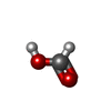

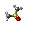

| #2: Chemical | 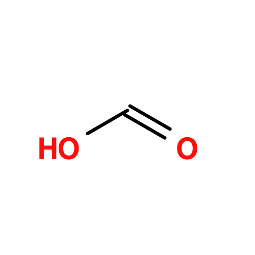 Mass: 46.025 Da / Num. of mol.: 2 / Source method: obtained synthetically / Formula: CH2O2 Mass: 46.025 Da / Num. of mol.: 2 / Source method: obtained synthetically / Formula: CH2O2#3: Chemical | ChemComp-A1J91 / | Mass: 266.293 Da / Num. of mol.: 1 / Source method: obtained synthetically / Formula: C13H18N2O4 #4: Chemical |  Mass: 24.305 Da / Num. of mol.: 2 / Source method: obtained synthetically / Formula: Mg Mass: 24.305 Da / Num. of mol.: 2 / Source method: obtained synthetically / Formula: Mg#5: Chemical | 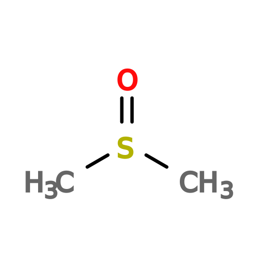 Mass: 78.133 Da / Num. of mol.: 2 / Source method: obtained synthetically / Formula: C2H6OS / Comment: DMSO, precipitant*YM Mass: 78.133 Da / Num. of mol.: 2 / Source method: obtained synthetically / Formula: C2H6OS / Comment: DMSO, precipitant*YM#6: Water | ChemComp-HOH / | Mass: 18.015 Da / Num. of mol.: 1367 / Source method: isolated from a natural source / Formula: H2O |
|---|
-Details
| Has protein modification | N |
|---|
-Experimental details
-Experiment
| Experiment | Method: X-RAY DIFFRACTION / Number of used crystals: 1 |
|---|
- Sample preparation
| Crystal | Density Matthews: 2.09 Å3/Da / Density % sol: 41.16 % |
|---|---|
| Crystal grow | Temperature: 293 K / Method: vapor diffusion, sitting drop Details: 20% (w/v) PEG 3350, 200 mM Magnesium formate, 100 mM HEPES pH 7.4 |
-Data collection
| Diffraction | Mean temperature: 100 K / Serial crystal experiment: N | ||||||||||||||||||||||||
|---|---|---|---|---|---|---|---|---|---|---|---|---|---|---|---|---|---|---|---|---|---|---|---|---|---|
| Diffraction source | Source: SYNCHROTRON / Site: PETRA III, EMBL c/o DESY  / Beamline: P14 (MX2) / Wavelength: 0.8265 Å / Beamline: P14 (MX2) / Wavelength: 0.8265 Å | ||||||||||||||||||||||||
| Detector | Type: DECTRIS EIGER2 X CdTe 16M / Detector: PIXEL / Date: Jan 26, 2026 | ||||||||||||||||||||||||
| Radiation | Protocol: SINGLE WAVELENGTH / Monochromatic (M) / Laue (L): M / Scattering type: x-ray | ||||||||||||||||||||||||
| Radiation wavelength | Wavelength: 0.8265 Å / Relative weight: 1 | ||||||||||||||||||||||||
| Reflection | Resolution: 1.005→49.744 Å / Num. obs: 230080 / % possible obs: 94 % / Redundancy: 8.8 % / Biso Wilson estimate: 9.96 Å2 / CC1/2: 0.999 / Rmerge(I) obs: 0.073 / Rpim(I) all: 0.038 / Rrim(I) all: 0.082 / Net I/σ(I): 14.1 | ||||||||||||||||||||||||
| Reflection shell | Num. unique obs: 11504 / Diffraction-ID: 1
|
- Processing
| Software |
| ||||||||||||||||||||||||||||||||||||||||||||||||||||||||||||||||||
|---|---|---|---|---|---|---|---|---|---|---|---|---|---|---|---|---|---|---|---|---|---|---|---|---|---|---|---|---|---|---|---|---|---|---|---|---|---|---|---|---|---|---|---|---|---|---|---|---|---|---|---|---|---|---|---|---|---|---|---|---|---|---|---|---|---|---|---|
| Refinement | Method to determine structure: MOLECULAR REPLACEMENT / Resolution: 1.2→17.54 Å / Cor.coef. Fo:Fc: 0.979 / Cor.coef. Fo:Fc free: 0.971 / SU R Cruickshank DPI: 0.039 / Cross valid method: THROUGHOUT / SU R Blow DPI: 0.038 / SU Rfree Blow DPI: 0.041 / SU Rfree Cruickshank DPI: 0.039
| ||||||||||||||||||||||||||||||||||||||||||||||||||||||||||||||||||
| Displacement parameters | Biso mean: 14.91 Å2
| ||||||||||||||||||||||||||||||||||||||||||||||||||||||||||||||||||
| Refine analyze | Luzzati coordinate error obs: 0.1 Å | ||||||||||||||||||||||||||||||||||||||||||||||||||||||||||||||||||
| Refinement step | Cycle: LAST / Resolution: 1.2→17.54 Å
| ||||||||||||||||||||||||||||||||||||||||||||||||||||||||||||||||||
| Refine LS restraints |
| ||||||||||||||||||||||||||||||||||||||||||||||||||||||||||||||||||
| LS refinement shell | Resolution: 1.2→1.22 Å
|
