 Movie
Movie Controller

[English] 日本語
 Yorodumi
- PDB-2zqo: Crystal structure of the earthworm R-type lectin C-half in comple... -
+ Open data

- Basic information
| Entry | Database: PDB / ID: 2zqo | |||||||||
|---|---|---|---|---|---|---|---|---|---|---|
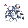
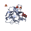
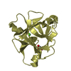
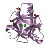
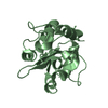
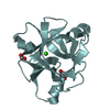
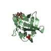
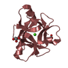
| Title | Crystal structure of the earthworm R-type lectin C-half in complex with GalNAc | |||||||||
 Components Components | 29-kDa galactose-binding lectin | |||||||||
 Keywords Keywords | SUGAR BINDING PROTEIN / EARTHWORM LUMBRICUS TERRESTRIS / HEMAGGLUTININ / R-TYPE LECTIN / BETA-TREFOIL FOLD / SUGAR COMPLEX / Lectin | |||||||||
| Function / homology |  Function and homology information Function and homology information | |||||||||
| Biological species |  Lumbricus terrestris (common earthworm) Lumbricus terrestris (common earthworm) | |||||||||
| Method |  X-RAY DIFFRACTION / SYNCHROTRON / MOLECULAR REPLACEMENT / Resolution: 1.8 Å X-RAY DIFFRACTION / SYNCHROTRON / MOLECULAR REPLACEMENT / Resolution: 1.8 Å | |||||||||
 Authors Authors | Suzuki, R. / Kuno, A. / Hasegawa, T. / Hirabayashi, J. / Kasai, K. / Momma, M. / Fujimoto, Z. | |||||||||
 Citation Citation | Journal: Acta Crystallogr.,Sect.D / Year: 2009 Title: Sugar-complex structures of the C-half domain of the galactose-binding lectin EW29 from the earthworm Lumbricus terrestris Authors: Suzuki, R. / Kuno, A. / Hasegawa, T. / Hirabayashi, J. / Kasai, K. / Momma, M. / Fujimoto, Z. #1: Journal: Acta Crystallogr.,Sect.D / Year: 2004 Title: Crystallization and preliminary X-ray crystallographic studies of the C-terminal domain of galactose-binding lectin EW29 from the earthworm Lumbricus terrestris Authors: Suzuki, R. / Fujimoto, Z. / Kuno, A. / Hirabayashi, J. / Kasai, K. / Hasegawa, T. #2: Journal: J.Biochem. / Year: 2007Title: Tailoring a novel sialic acid-binding lectin from a ricin-B chain-like galactose-binding protein by natural evolution-mimicry Authors: Yabe, R. / Suzuki, R. / Kuno, A. / Fujimoto, Z. / Jigami, Y. / Hirabayashi, J. #3: Journal: J.Biol.Chem. / Year: 1998 Title: Novel galactose-binding proteins in Annelida. Characterization of 29-kDa tandem repeat-type lectins from the earthworm Lumbricus terrestris Authors: Hirabayashi, J. / Dutta, S.K. / Kasai, K. | |||||||||
| History |
|
- Structure visualization
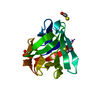
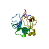
| Structure viewer | Molecule: MolmilJmol/JSmol |
|---|
- Downloads & links
-Download
| PDBx/mmCIF format | 2zqo.cif.gz | 74.6 KB | Display | PDBx/mmCIF format |
|---|---|---|---|---|
| PDB format | pdb2zqo.ent.gz | 55.2 KB | Display | PDB format |
| PDBx/mmJSON format | 2zqo.json.gz | Tree view | PDBx/mmJSON format | |
| Others |  Other downloads Other downloads |
-Validation report
| Arichive directory | https://data.pdbj.org/pub/pdb/validation_reports/zq/2zqoftp://data.pdbj.org/pub/pdb/validation_reports/zq/2zqo | HTTPS FTP |
|---|
-Related structure data
| Related structure data |  2zqnC 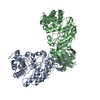 1xyfS C: citing same article ( S: Starting model for refinement |
|---|---|
| Similar structure data |


-Links
 PDBj
PDBj


- Assembly
| Deposited unit | 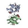
| ||||||||
|---|---|---|---|---|---|---|---|---|---|
| 1 | 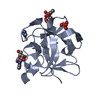
| ||||||||
| 2 | 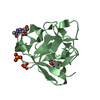
| ||||||||
| Unit cell |
| ||||||||
| Components on special symmetry positions |
|
-Components
-Protein / Sugars , 2 types, 6 molecules AB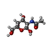
| #1: Protein | Mass: 14617.369 Da / Num. of mol.: 2 / Fragment: C-terminal domain Source method: isolated from a genetically manipulated source Source: (gene. exp.) Lumbricus terrestris (common earthworm)Plasmid: pET21 / Production host:  #2: Sugar | ChemComp-NGA /  Type: D-saccharide, beta linking / Mass: 221.208 Da / Num. of mol.: 4 Type: D-saccharide, beta linking / Mass: 221.208 Da / Num. of mol.: 4Source method: isolated from a genetically manipulated source Formula: C8H15NO6 |
|---|
-Non-polymers , 4 types, 320 molecules 


| #3: Chemical | ChemComp-CD /  Mass: 112.411 Da / Num. of mol.: 1 / Source method: obtained synthetically / Formula: Cd Mass: 112.411 Da / Num. of mol.: 1 / Source method: obtained synthetically / Formula: Cd | ||||
|---|---|---|---|---|---|
| #4: Chemical |  Mass: 94.971 Da / Num. of mol.: 3 / Source method: obtained synthetically / Formula: PO4 Mass: 94.971 Da / Num. of mol.: 3 / Source method: obtained synthetically / Formula: PO4#5: Chemical | ChemComp-IMD / |  Mass: 69.085 Da / Num. of mol.: 1 / Source method: obtained synthetically / Formula: C3H5N2 Mass: 69.085 Da / Num. of mol.: 1 / Source method: obtained synthetically / Formula: C3H5N2#6: Water | ChemComp-HOH / | Mass: 18.015 Da / Num. of mol.: 315 / Source method: isolated from a natural source / Formula: H2O |
-Experimental details
-Experiment
| Experiment | Method: X-RAY DIFFRACTION / Number of used crystals: 1 |
|---|
- Sample preparation
| Crystal | Density Matthews: 2.19 Å3/Da / Density % sol: 43.73 % |
|---|---|
| Crystal grow | Temperature: 293 K / Method: vapor diffusion, sitting drop / pH: 8 Details: sodium chloride, dipotassium hydrogen phosphate, sodium dihydrogen phosphate, imidazole, cadmium chloride, pH 8.0, VAPOR DIFFUSION, SITTING DROP, temperature 293K |
-Data collection
| Diffraction | Mean temperature: 95 K |
|---|---|
| Diffraction source | Source: SYNCHROTRON / Site: Photon Factory  / Beamline: BL-6A / Wavelength: 1 Å / Beamline: BL-6A / Wavelength: 1 Å |
| Detector | Type: ADSC QUANTUM 4 / Detector: CCD / Date: Nov 5, 2004 |
| Radiation | Protocol: SINGLE WAVELENGTH / Monochromatic (M) / Laue (L): M / Scattering type: x-ray |
| Radiation wavelength | Wavelength: 1 Å / Relative weight: 1 |
| Reflection | Resolution: 1.8→50 Å / Num. obs: 23141 / % possible obs: 94 % / Observed criterion σ(I): 0 / Redundancy: 7.2 % / Rmerge(I) obs: 0.098 / Net I/σ(I): 36.1 |
| Reflection shell | Resolution: 1.8→1.86 Å / Redundancy: 7 % / Rmerge(I) obs: 0.308 / Mean I/σ(I) obs: 8.6 / Num. unique all: 2213 / % possible all: 94 |
- Processing
| Software |
| ||||||||||||||||||||||||||||||||||||||||||||||||||||||||||||||||||||||||||||||||||||||||||
|---|---|---|---|---|---|---|---|---|---|---|---|---|---|---|---|---|---|---|---|---|---|---|---|---|---|---|---|---|---|---|---|---|---|---|---|---|---|---|---|---|---|---|---|---|---|---|---|---|---|---|---|---|---|---|---|---|---|---|---|---|---|---|---|---|---|---|---|---|---|---|---|---|---|---|---|---|---|---|---|---|---|---|---|---|---|---|---|---|---|---|---|
| Refinement | Method to determine structure: MOLECULAR REPLACEMENT Starting model: PDB ENTRY 1XYF Resolution: 1.8→48.62 Å / Cor.coef. Fo:Fc: 0.939 / Cor.coef. Fo:Fc free: 0.9 / SU B: 2.766 / SU ML: 0.09 / Isotropic thermal model: RESTRAINED / Cross valid method: THROUGHOUT / σ(F): 0 / ESU R: 0.161 / ESU R Free: 0.161 / Stereochemistry target values: MAXIMUM LIKELIHOOD / Details: HYDROGENS HAVE BEEN ADDED IN THE RIDING POSITIONS
| ||||||||||||||||||||||||||||||||||||||||||||||||||||||||||||||||||||||||||||||||||||||||||
| Solvent computation | Ion probe radii: 0.8 Å / Shrinkage radii: 0.8 Å / VDW probe radii: 1.2 Å / Solvent model: MASK | ||||||||||||||||||||||||||||||||||||||||||||||||||||||||||||||||||||||||||||||||||||||||||
| Displacement parameters | Biso mean: 15.126 Å2
| ||||||||||||||||||||||||||||||||||||||||||||||||||||||||||||||||||||||||||||||||||||||||||
| Refinement step | Cycle: LAST / Resolution: 1.8→48.62 Å
| ||||||||||||||||||||||||||||||||||||||||||||||||||||||||||||||||||||||||||||||||||||||||||
| Refine LS restraints |
| ||||||||||||||||||||||||||||||||||||||||||||||||||||||||||||||||||||||||||||||||||||||||||
| LS refinement shell | Resolution: 1.8→1.847 Å / Total num. of bins used: 20
|
