 Movie
Movie Controller

[English] 日本語
 Yorodumi
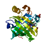
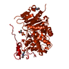
- PDB-2zm2: Structure of 6-aminohexanoate-dimer hydrolase, A61V/A124V/R187S/F... -
+ Open data

- Basic information
| Entry | Database: PDB / ID: 2zm2 | ||||||
|---|---|---|---|---|---|---|---|
| Title | Structure of 6-aminohexanoate-dimer hydrolase, A61V/A124V/R187S/F264C/G291R/G338A/D370Y mutant (Hyb-S4M94) | ||||||
 Components Components | 6-aminohexanoate-dimer hydrolase | ||||||
 Keywords Keywords | HYDROLASE / ALPHA-BETA | ||||||
| Function / homology |  Function and homology information Function and homology information6-aminohexanoate-oligomer exohydrolase / 6-aminohexanoate-dimer hydrolase activity / nylon catabolic process Similarity search - Function | ||||||
| Biological species |  FLAVOBACTERIUM SP. (bacteria) FLAVOBACTERIUM SP. (bacteria) | ||||||
| Method |  X-RAY DIFFRACTION / SYNCHROTRON / MOLECULAR REPLACEMENT / Resolution: 1.55 Å X-RAY DIFFRACTION / SYNCHROTRON / MOLECULAR REPLACEMENT / Resolution: 1.55 Å | ||||||
 Authors Authors | Ohki, T. / Shibata, N. / Higuchi, Y. / Kawashima, Y. / Takeo, M. / Kato, D. / Nego, S. | ||||||
 Citation Citation | Journal: Protein Sci. / Year: 2009 Title: Two alternative modes for optimizing nylon-6 byproduct hydrolytic activity from a carboxylesterase with a beta-lactamase fold: X-ray crystallographic analysis of directly evolved 6-aminohexanoate-dimer hydrolase. Authors: Ohki, T. / Shibata, N. / Higuchi, Y. / Kawashima, Y. / Takeo, M. / Kato, D. / Negoro, S. | ||||||
| History |
|
- Structure visualization
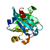
| Structure viewer | Molecule: MolmilJmol/JSmol |
|---|
- Downloads & links
-Download
| PDBx/mmCIF format | 2zm2.cif.gz | 103.3 KB | Display | PDBx/mmCIF format |
|---|---|---|---|---|
| PDB format | pdb2zm2.ent.gz | 76.5 KB | Display | PDB format |
| PDBx/mmJSON format | 2zm2.json.gz | Tree view | PDBx/mmJSON format | |
| Others |  Other downloads Other downloads |
-Validation report
| Arichive directory | https://data.pdbj.org/pub/pdb/validation_reports/zm/2zm2ftp://data.pdbj.org/pub/pdb/validation_reports/zm/2zm2 | HTTPS FTP |
|---|
-Related structure data
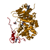
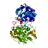
| Related structure data |  2zlyC  2zm8C  2zm9C  1wybS C: citing same article ( S: Starting model for refinement |
|---|---|
| Similar structure data |


-Links
 PDBj
PDBj
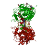
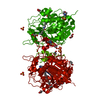
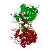
- Assembly
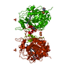
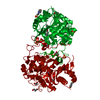
| Deposited unit | 
| |||||||||
|---|---|---|---|---|---|---|---|---|---|---|
| 1 | 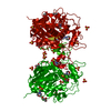
| |||||||||
| 2 |
| |||||||||
| Unit cell |
| |||||||||
| Components on special symmetry positions |
|
-Components
| #1: Protein | Mass: 42943.711 Da / Num. of mol.: 1 / Mutation: A61V, A124V, R187S, F264C, G291R, G338A, D370Y Source method: isolated from a genetically manipulated source Details: CHIMERA OF NYLON OLIGOMERS-DEGRADING ENZYME EII (RESIDUES 1-21) AND NYLON OLIGOMERS-DEGRADING ENZYME EII' (RESIDUES 22-392) Source: (gene. exp.) FLAVOBACTERIUM SP. (bacteria) / Strain: K172 / Gene: NYLB, NYLB' / Plasmid: PKP1500 / Production host: References: UniProt: P07061, UniProt: P07062, 6-aminohexanoate-oligomer exohydrolase | ||||||||
|---|---|---|---|---|---|---|---|---|---|
| #2: Chemical | ChemComp-SO4 /   Mass: 96.063 Da / Num. of mol.: 5 / Source method: obtained synthetically / Formula: SO4 Mass: 96.063 Da / Num. of mol.: 5 / Source method: obtained synthetically / Formula: SO4#3: Chemical | ChemComp-MES /   Mass: 195.237 Da / Num. of mol.: 4 / Source method: obtained synthetically / Formula: C6H13NO4S / Comment: pH buffer*YM Mass: 195.237 Da / Num. of mol.: 4 / Source method: obtained synthetically / Formula: C6H13NO4S / Comment: pH buffer*YM#4: Chemical | ChemComp-GOL /   Mass: 92.094 Da / Num. of mol.: 4 / Source method: obtained synthetically / Formula: C3H8O3 Mass: 92.094 Da / Num. of mol.: 4 / Source method: obtained synthetically / Formula: C3H8O3#5: Water | ChemComp-HOH / |  Mass: 18.015 Da / Num. of mol.: 495 / Source method: isolated from a natural source / Formula: H2O Mass: 18.015 Da / Num. of mol.: 495 / Source method: isolated from a natural source / Formula: H2OSequence details | ACCORDING TO DEPOSITORS | |
-Experimental details
-Experiment
| Experiment | Method: X-RAY DIFFRACTION / Number of used crystals: 1 |
|---|
- Sample preparation
| Crystal | Density Matthews: 3.55 Å3/Da / Density % sol: 65.33 % |
|---|---|
| Crystal grow | Temperature: 283 K / pH: 6.5 Details: 2.2M ammonium sulfate, 0.2M lithium sulfate, 0.1M MES, pH 6.50, temperature 283K |
-Data collection
| Diffraction | Mean temperature: 100 K |
|---|---|
| Diffraction source | Source: SYNCHROTRON / Site: SPring-8  / Beamline: BL38B1 / Wavelength: 0.9 / Wavelength: 0.9 Å / Beamline: BL38B1 / Wavelength: 0.9 / Wavelength: 0.9 Å |
| Detector | Type: RIGAKU JUPITER 210 / Detector: CCD / Date: Jun 18, 2005 |
| Radiation | Protocol: SINGLE WAVELENGTH / Monochromatic (M) / Laue (L): M / Scattering type: x-ray |
| Radiation wavelength | Wavelength: 0.9 Å / Relative weight: 1 |
| Reflection | Resolution: 1.5→50 Å / Num. all: 96878 / Num. obs: 96878 / % possible obs: 100 % / Observed criterion σ(I): -3 / Redundancy: 10.9 % / Biso Wilson estimate: 17.2 Å2 / Rmerge(I) obs: 0.125 / Net I/σ(I): 44.91 |
| Reflection shell | Resolution: 1.55→1.61 Å / Redundancy: 10.6 % / Rmerge(I) obs: 0.446 / Mean I/σ(I) obs: 4.26 / Num. unique all: 8782 / % possible all: 100 |
- Processing
| Software |
| ||||||||||||||||||||||||||||||||||||||||||||||||||||||||||||||||||||||||||||||||
|---|---|---|---|---|---|---|---|---|---|---|---|---|---|---|---|---|---|---|---|---|---|---|---|---|---|---|---|---|---|---|---|---|---|---|---|---|---|---|---|---|---|---|---|---|---|---|---|---|---|---|---|---|---|---|---|---|---|---|---|---|---|---|---|---|---|---|---|---|---|---|---|---|---|---|---|---|---|---|---|---|---|
| Refinement | Method to determine structure: MOLECULAR REPLACEMENT Starting model: PDB ENTRY 1WYB Resolution: 1.55→33.64 Å / Rfactor Rfree error: 0.002 / Data cutoff high absF: 1845983.03 / Data cutoff low absF: 0 / Isotropic thermal model: RESTRAINED / Cross valid method: THROUGHOUT / σ(F): 0 / Stereochemistry target values: Engh & Huber
| ||||||||||||||||||||||||||||||||||||||||||||||||||||||||||||||||||||||||||||||||
| Solvent computation | Solvent model: FLAT MODEL / Bsol: 62.0769 Å2 / ksol: 0.387215 e/Å3 | ||||||||||||||||||||||||||||||||||||||||||||||||||||||||||||||||||||||||||||||||
| Displacement parameters | Biso mean: 20.1 Å2
| ||||||||||||||||||||||||||||||||||||||||||||||||||||||||||||||||||||||||||||||||
| Refine analyze |
| ||||||||||||||||||||||||||||||||||||||||||||||||||||||||||||||||||||||||||||||||
| Refinement step | Cycle: LAST / Resolution: 1.55→33.64 Å
| ||||||||||||||||||||||||||||||||||||||||||||||||||||||||||||||||||||||||||||||||
| Refine LS restraints |
| ||||||||||||||||||||||||||||||||||||||||||||||||||||||||||||||||||||||||||||||||
| LS refinement shell | Resolution: 1.55→1.61 Å / Rfactor Rfree error: 0.008 / Total num. of bins used: 10
|
