[English] 日本語
Yorodumi
- PDB-2zfo: Structure of the partially unliganded met state of 400 kDa hemogl... -

+
Open data


ID or keywords:

Loading...

-
Basic information

Entry
Database: PDB / ID: 2zfo
TitleStructure of the partially unliganded met state of 400 kDa hemoglobin: Insights into ligand-induced structural changes of giant hemoglobins
Components(Extracellular giant hemoglobin major globin subunit ...) x 4
KeywordsOXYGEN BINDING / TRANSPORT PROTEIN / hemoglobin / polychaete / annelida / unliganded / Heme / Iron / Metal-binding / Oxygen transport / Secreted / Transport
Function / homology
Function and homology information


hemoglobin complex / oxygen carrier activity / oxygen binding / response to hypoxia / iron ion binding / heme binding / extracellular region
Similarity search - Function
Globin, extracellular / Erythrocruorin / Myoglobin-like, M family globin domain / Globin/Protoglobin / Globins / Globin domain profile. / Globin-like / Globin / Globin / Globin-like superfamily ...Globin, extracellular / Erythrocruorin / Myoglobin-like, M family globin domain / Globin/Protoglobin / Globins / Globin domain profile. / Globin-like / Globin / Globin / Globin-like superfamily / Orthogonal Bundle / Mainly Alpha
Similarity search - Domain/homology
PROTOPORPHYRIN IX CONTAINING FE / OXYGEN MOLECULE / Extracellular giant hemoglobin major globin subunit B1 / Extracellular giant hemoglobin major globin subunit A2 / Extracellular giant hemoglobin major globin subunit B2 / Extracellular giant hemoglobin major globin subunit A1
Similarity search - Component
Biological speciesOligobrachia mashikoi (invertebrata)
MethodX-RAY DIFFRACTION / SYNCHROTRON / MOLECULAR REPLACEMENT / Resolution: 1.95 Å
AuthorsNumoto, N. / Nakagawa, T. / Kita, A. / Sasayama, Y. / Fukumori, Y. / Miki, K.
CitationJournal: Proteins / Year: 2008
Title: Structure of the partially unliganded met state of 400 kDa hemoglobin: insights into ligand-induced structural changes of giant hemoglobins
Authors: Numoto, N. / Nakagawa, T. / Kita, A. / Sasayama, Y. / Fukumori, Y. / Miki, K.
History
DepositionJan 8, 2008Deposition site: PDBJ / Processing site: PDBJ
Revision 1.0Apr 22, 2008Provider: repository / Type: Initial release
Revision 1.1Jul 13, 2011Group: Non-polymer description / Version format compliance
Revision 1.2Nov 1, 2023Group: Data collection / Database references ...Data collection / Database references / Derived calculations / Refinement description
Category: chem_comp_atom / chem_comp_bond ...chem_comp_atom / chem_comp_bond / database_2 / pdbx_initial_refinement_model / struct_conn / struct_ref_seq_dif / struct_site
Item: _database_2.pdbx_DOI / _database_2.pdbx_database_accession ..._database_2.pdbx_DOI / _database_2.pdbx_database_accession / _struct_conn.pdbx_dist_value / _struct_conn.ptnr1_auth_asym_id / _struct_conn.ptnr1_auth_comp_id / _struct_conn.ptnr1_auth_seq_id / _struct_conn.ptnr1_label_asym_id / _struct_conn.ptnr1_label_atom_id / _struct_conn.ptnr1_label_comp_id / _struct_conn.ptnr1_label_seq_id / _struct_conn.ptnr2_auth_asym_id / _struct_conn.ptnr2_auth_comp_id / _struct_conn.ptnr2_auth_seq_id / _struct_conn.ptnr2_label_asym_id / _struct_conn.ptnr2_label_atom_id / _struct_conn.ptnr2_label_comp_id / _struct_ref_seq_dif.details / _struct_site.pdbx_auth_asym_id / _struct_site.pdbx_auth_comp_id / _struct_site.pdbx_auth_seq_id
Remark 650HELIX DETERMINATION METHOD: AUTHOR DETERMINED

-
Structure visualization

Structure viewerMolecule:
MolmilJmol/JSmol

Downloads & links

-
Assembly

Deposited unit
A: Extracellular giant hemoglobin major globin subunit A1
B: Extracellular giant hemoglobin major globin subunit A2
C: Extracellular giant hemoglobin major globin subunit B2
D: Extracellular giant hemoglobin major globin subunit B1
hetero molecules


Theoretical massNumber of molelcules
Total (without water)63,51311
Polymers60,8314
Non-polymers2,6827
Water9,422523
1
A: Extracellular giant hemoglobin major globin subunit A1
B: Extracellular giant hemoglobin major globin subunit A2
C: Extracellular giant hemoglobin major globin subunit B2
D: Extracellular giant hemoglobin major globin subunit B1
hetero molecules
x 6


Theoretical massNumber of molelcules
Total (without water)381,07666
Polymers364,98424
Non-polymers16,09342
Water43224
TypeNameSymmetry operationNumber
identity operation1_555x,y,z1
crystal symmetry operation2_555-y,x-y,z1
crystal symmetry operation3_555-x+y,-x,z1
crystal symmetry operation4_555y,x,-z1
crystal symmetry operation5_555x-y,-y,-z1
crystal symmetry operation6_555-x,-x+y,-z1
Buried area91850 Å2
ΔGint-731 kcal/mol
Surface area110740 Å2
MethodPISA
Unit cell
Length a, b, c (Å)110.960, 110.960, 271.580
Angle α, β, γ (deg.)90.00, 90.00, 120.00
Int Tables number155
Space group name H-MH32
Components on special symmetry positions
IDModelComponents
11A-1269-

HOH

21C-1277-

HOH

-
Components

-
Extracellular giant hemoglobin major globin subunit ... , 4 types, 4 molecules ABCD

#1: Protein Extracellular giant hemoglobin major globin subunit A1 / Major globin chain b


Mass: 15188.894 Da / Num. of mol.: 1 / Source method: isolated from a natural source / Source: (natural) Oligobrachia mashikoi (invertebrata) / References: UniProt: Q7M419
#2: Protein Extracellular giant hemoglobin major globin subunit A2 / Major globin chain a5


Mass: 15327.087 Da / Num. of mol.: 1 / Source method: isolated from a natural source / Source: (natural) Oligobrachia mashikoi (invertebrata) / References: UniProt: Q7M413
#3: Protein Extracellular giant hemoglobin major globin subunit B2 / Major globin chain c


Mass: 15621.285 Da / Num. of mol.: 1 / Source method: isolated from a natural source / Source: (natural) Oligobrachia mashikoi (invertebrata) / References: UniProt: Q7M418
#4: Protein Extracellular giant hemoglobin major globin subunit B1 / Major globin chain d


Mass: 14693.341 Da / Num. of mol.: 1 / Source method: isolated from a natural source / Source: (natural) Oligobrachia mashikoi (invertebrata) / References: UniProt: Q5KSB7

-
Non-polymers , 4 types, 530 molecules

#5: Chemical
ChemComp-HEM / PROTOPORPHYRIN IX CONTAINING FE / HEME


Mass: 616.487 Da / Num. of mol.: 4 / Source method: obtained synthetically / Formula: C34H32FeN4O4
#6: Chemical ChemComp-OXY / OXYGEN MOLECULE


Mass: 31.999 Da / Num. of mol.: 1 / Source method: obtained synthetically / Formula: O2
#7: Chemical ChemComp-GOL / GLYCEROL / GLYCERIN / PROPANE-1,2,3-TRIOL


Mass: 92.094 Da / Num. of mol.: 2 / Source method: obtained synthetically / Formula: C3H8O3
#8: Water ChemComp-HOH / water


Mass: 18.015 Da / Num. of mol.: 523 / Source method: isolated from a natural source / Formula: H2O

-
Details

Sequence details
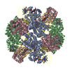
THE CHAIN D (SUBUNIT B1) IS THE MIXTURE OF TWO ISOFORMS (B1A AND B1B) CONTAINING 10 SUBSTITUTED ...THE CHAIN D (SUBUNIT B1) IS THE MIXTURE OF TWO ISOFORMS (B1A AND B1B) CONTAINING 10 SUBSTITUTED RESIDUES IN EACH 145 AMINO ACIDS. WE CONSTRUCTED THE MODEL BASICALLY BY USING THE SEQUENCE OF B1A AND INCLUDED THE MODELS OF THE 10 SUBSTITUTED RESIDUES OF THE B1B AS PSEUDO DOUBLE CONFORMATIONS, BECAUSE INCLUDING OF THE FULL LENGTH OF BOTH B1A AND B1B WITH A 50/50 DISTRIBUTION WILL RESULTS IN UNDESIRED INCREASES IN THE PARAMETERS.

-
Experimental details

-
Experiment

ExperimentMethod: X-RAY DIFFRACTION / Number of used crystals: 1

-
Sample preparation

CrystalDensity Matthews: 2.64 Å3/Da / Density % sol: 53.49 %
Crystal growTemperature: 293 K / Method: vapor diffusion, hanging drop / pH: 8
Details: 7% PEG 10000, 0.2M Tris-HCl (pH 8.0), VAPOR DIFFUSION, HANGING DROP, temperature 293K

-
Data collection

DiffractionMean temperature: 90 K
Diffraction sourceSource: SYNCHROTRON / Site: SPring-8 / Beamline: BL26B1 / Wavelength: 1 Å
DetectorType: RIGAKU JUPITER 210 / Detector: CCD / Date: Jun 29, 2006 / Details: Rh-coated cylindrical bending mirror
RadiationMonochromator: Fixed exit Si 111 double-crystal monochromator
Protocol: SINGLE WAVELENGTH / Monochromatic (M) / Laue (L): M / Scattering type: x-ray
Radiation wavelengthWavelength: 1 Å / Relative weight: 1
ReflectionResolution: 1.95→50 Å / Num. all: 47042 / Num. obs: 47042 / % possible obs: 99.7 % / Observed criterion σ(I): -3 / Redundancy: 9.2 % / Biso Wilson estimate: 22.9 Å2 / Rsym value: 0.061 / Net I/σ(I): 32.8
Reflection shellResolution: 1.95→2.02 Å / Redundancy: 7.2 % / Mean I/σ(I) obs: 5.8 / Num. unique all: 4642 / Rsym value: 0.337 / % possible all: 99.4

-
Processing

Software
NameVersionClassification
CNS1.2refinement
BSSdata collection
HKL-2000data reduction
SCALEPACKdata scaling
MOLREPphasing
RefinementMethod to determine structure: MOLECULAR REPLACEMENT
Starting model: PDB ENTRY 2D2M
Resolution: 1.95→39.22 Å / Rfactor Rfree error: 0.004 / Data cutoff high absF: 4031385.32 / Data cutoff low absF: 0 / Isotropic thermal model: RESTRAINED / Cross valid method: THROUGHOUT / σ(F): 0 / Stereochemistry target values: Engh & Huber / Details: BULK SOLVENT MODEL USED
RfactorNum. reflection% reflectionSelection details
Rfree0.202 2332 5 %RANDOM
Rwork0.169 ---
obs-46892 99.2 %-
Solvent computationSolvent model: FLAT MODEL / Bsol: 70.2087 Å2 / ksol: 0.4 e/Å3
Displacement parametersBiso mean: 29.5 Å2
Baniso -1Baniso -2Baniso -3
1-4.57 Å20 Å20 Å2
2--4.57 Å20 Å2
3----9.14 Å2
Refine analyze
FreeObs
Luzzati coordinate error0.23 Å0.18 Å
Luzzati d res low-5 Å
Luzzati sigma a0.19 Å0.14 Å
Refinement stepCycle: LAST / Resolution: 1.95→39.22 Å
ProteinNucleic acidLigandSolventTotal
Num. atoms4339 0 186 523 5048
Refine LS restraints
Refine-IDTypeDev idealDev ideal target
X-RAY DIFFRACTIONc_bond_d0.01
X-RAY DIFFRACTIONc_bond_d_na
X-RAY DIFFRACTIONc_bond_d_prot
X-RAY DIFFRACTIONc_angle_d
X-RAY DIFFRACTIONc_angle_d_na
X-RAY DIFFRACTIONc_angle_d_prot
X-RAY DIFFRACTIONc_angle_deg1.3
X-RAY DIFFRACTIONc_angle_deg_na
X-RAY DIFFRACTIONc_angle_deg_prot
X-RAY DIFFRACTIONc_dihedral_angle_d18.6
X-RAY DIFFRACTIONc_dihedral_angle_d_na
X-RAY DIFFRACTIONc_dihedral_angle_d_prot
X-RAY DIFFRACTIONc_improper_angle_d0.95
X-RAY DIFFRACTIONc_improper_angle_d_na
X-RAY DIFFRACTIONc_improper_angle_d_prot
X-RAY DIFFRACTIONc_mcbond_it1.261.5
X-RAY DIFFRACTIONc_mcangle_it1.842
X-RAY DIFFRACTIONc_scbond_it2.032
X-RAY DIFFRACTIONc_scangle_it2.842.5
LS refinement shellResolution: 1.95→2.02 Å / Rfactor Rfree error: 0.017 / Total num. of bins used: 10
RfactorNum. reflection% reflection
Rfree0.26 230 5 %
Rwork0.22 4340 -
obs-2332 97.8 %
Xplor file
Refine-IDSerial noParam fileTopol file
X-RAY DIFFRACTION1protein_rep.paramprotein.top
X-RAY DIFFRACTION2water_rep.paramwater.top
X-RAY DIFFRACTION3hem_o2_final.paramhem_o2_final.top
X-RAY DIFFRACTION4gol_final.paramgol_final.par

+
About Yorodumi

-
News

-
Feb 9, 2022. New format data for meta-information of EMDB entries

New format data for meta-information of EMDB entries

  • Version 3 of the EMDB header file is now the official format.
  • The previous official version 1.9 will be removed from the archive.

Related info.:EMDB header

External links:wwPDB to switch to version 3 of the EMDB data model

-
Aug 12, 2020. Covid-19 info

Covid-19 info

URL: https://pdbjlvh1.pdbj.org/emnavi/covid19.php

New page: Covid-19 featured information page in EM Navigator.

Related info.:Covid-19 info / Mar 5, 2020. Novel coronavirus structure data

+
Mar 5, 2020. Novel coronavirus structure data

Novel coronavirus structure data

Related info.:Yorodumi Speices / Aug 12, 2020. Covid-19 info

External links:COVID-19 featured content - PDBj / Molecule of the Month (242):Coronavirus Proteases

+
Jan 31, 2019. EMDB accession codes are about to change! (news from PDBe EMDB page)

EMDB accession codes are about to change! (news from PDBe EMDB page)

  • The allocation of 4 digits for EMDB accession codes will soon come to an end. Whilst these codes will remain in use, new EMDB accession codes will include an additional digit and will expand incrementally as the available range of codes is exhausted. The current 4-digit format prefixed with “EMD-” (i.e. EMD-XXXX) will advance to a 5-digit format (i.e. EMD-XXXXX), and so on. It is currently estimated that the 4-digit codes will be depleted around Spring 2019, at which point the 5-digit format will come into force.
  • The EM Navigator/Yorodumi systems omit the EMD- prefix.

Related info.:Q: What is EMD? / ID/Accession-code notation in Yorodumi/EM Navigator

External links:EMDB Accession Codes are Changing Soon! / Contact to PDBj

+
Jul 12, 2017. Major update of PDB

Major update of PDB

  • wwPDB released updated PDB data conforming to the new PDBx/mmCIF dictionary.
  • This is a major update changing the version number from 4 to 5, and with Remediation, in which all the entries are updated.
  • In this update, many items about electron microscopy experimental information are reorganized (e.g. em_software).
  • Now, EM Navigator and Yorodumi are based on the updated data.

External links:wwPDB Remediation / Enriched Model Files Conforming to OneDep Data Standards Now Available in the PDB FTP Archive

-
Yorodumi

Thousand views of thousand structures

  • Yorodumi is a browser for structure data from EMDB, PDB, SASBDB, etc.
  • This page is also the successor to EM Navigator detail page, and also detail information page/front-end page for Omokage search.
  • The word "yorodu" (or yorozu) is an old Japanese word meaning "ten thousand". "mi" (miru) is to see.

Related info.:EMDB / PDB / SASBDB / Comparison of 3 databanks / Yorodumi Search / Aug 31, 2016. New EM Navigator & Yorodumi / Yorodumi Papers / Jmol/JSmol / Function and homology information / Changes in new EM Navigator and Yorodumi

Read more