 Movie
Movie Controller


+ Open data

- Basic information
| Entry | Database: PDB / ID: 2zbn | ||||||
|---|---|---|---|---|---|---|---|
| Title | Crystal structure of PH1033 from Pyrococcus horikoshii OT3 | ||||||
 Components Components | UPF0310 protein PH1033 | ||||||
 Keywords Keywords | STRUCTURAL GENOMICS / UNKNOWN FUNCTION / Pyrococcus horikoshii OT3 / NPPSFA / National Project on Protein Structural and Functional Analyses / RIKEN Structural Genomics/Proteomics Initiative / RSGI | ||||||
| Function / homology | Uncharacterised protein family UPF0310 / EVE domain / EVE domain / ph1033 like fold / ph1033 like domains / PUA-like superfamily / Roll / Alpha Beta / UPF0310 protein PH1033 Function and homology information Function and homology information | ||||||
| Biological species |   Pyrococcus horikoshii (archaea) Pyrococcus horikoshii (archaea) | ||||||
| Method |  X-RAY DIFFRACTION / SYNCHROTRON / MOLECULAR REPLACEMENT / Resolution: 2 Å X-RAY DIFFRACTION / SYNCHROTRON / MOLECULAR REPLACEMENT / Resolution: 2 Å | ||||||
 Authors Authors | Sugahara, M. / Kunishima, N. / RIKEN Structural Genomics/Proteomics Initiative (RSGI) | ||||||
 Citation Citation | Journal: Acta Crystallogr.,Sect.D / Year: 2008 Title: Nucleant-mediated protein crystallization with the application of microporous synthetic zeolites. Authors: Sugahara, M. / Asada, Y. / Morikawa, Y. / Kageyama, Y. / Kunishima, N. | ||||||
| History |
|
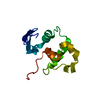
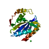
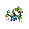
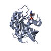
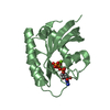
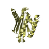
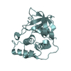
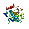
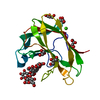
- Structure visualization
| Structure viewer | Molecule: MolmilJmol/JSmol |
|---|
- Downloads & links
-Download
| PDBx/mmCIF format | 2zbn.cif.gz | 44.9 KB | Display | PDBx/mmCIF format |
|---|---|---|---|---|
| PDB format | pdb2zbn.ent.gz | 31.2 KB | Display | PDB format |
| PDBx/mmJSON format | 2zbn.json.gz | Tree view | PDBx/mmJSON format | |
| Others |  Other downloads Other downloads |
-Validation report
| Arichive directory | https://data.pdbj.org/pub/pdb/validation_reports/zb/2zbnftp://data.pdbj.org/pub/pdb/validation_reports/zb/2zbn | HTTPS FTP |
|---|
-Related structure data
| Related structure data |  1wmmSC 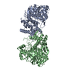 2dpnC 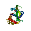 2hd9C S: Starting model for refinement C: citing same article ( |
|---|---|
| Similar structure data |

-Links
 PDBj
PDBj
- Assembly
| Deposited unit | 
| ||||||||
|---|---|---|---|---|---|---|---|---|---|
| 1 |
| ||||||||
| Unit cell |
| ||||||||
| Components on special symmetry positions |
|
-Components
| #1: Protein | Mass: 17464.623 Da / Num. of mol.: 1 Source method: isolated from a genetically manipulated source Source: (gene. exp.) Pyrococcus horikoshii (archaea) / Strain: OT3 / Plasmid: pET-11a / Production host:  |
|---|---|
| #2: Water | ChemComp-HOH /  Mass: 18.015 Da / Num. of mol.: 97 / Source method: isolated from a natural source / Formula: H2O Mass: 18.015 Da / Num. of mol.: 97 / Source method: isolated from a natural source / Formula: H2O |
-Experimental details
-Experiment
| Experiment | Method: X-RAY DIFFRACTION / Number of used crystals: 1 |
|---|
- Sample preparation
| Crystal | Density Matthews: 3.32 Å3/Da / Density % sol: 63 % |
|---|---|
| Crystal grow | Temperature: 295 K / pH: 5.7 Details: 22.5% PEG 4K, 15% Glycerol, 0.1M citrate buffer, pH 5.7, oil microbatch, temperature 295K |
-Data collection
| Diffraction | Mean temperature: 100 K |
|---|---|
| Diffraction source | Source: SYNCHROTRON / Site: SPring-8  / Beamline: BL26B1 / Wavelength: 1 Å / Beamline: BL26B1 / Wavelength: 1 Å |
| Detector | Type: RIGAKU RAXIS V / Detector: IMAGE PLATE / Date: Jul 22, 2005 |
| Radiation | Protocol: SINGLE WAVELENGTH / Monochromatic (M) / Laue (L): M / Scattering type: x-ray |
| Radiation wavelength | Wavelength: 1 Å / Relative weight: 1 |
| Reflection | Resolution: 2→40 Å / Num. obs: 15961 / % possible obs: 100 % / Observed criterion σ(F): 0 / Observed criterion σ(I): 0 / Redundancy: 9.6 % / Biso Wilson estimate: 33.4 Å2 / Rmerge(I) obs: 0.068 / Rsym value: 0.065 / Net I/σ(I): 9.9 |
| Reflection shell | Resolution: 2→2.07 Å / Redundancy: 9.4 % / Rmerge(I) obs: 0.585 / Mean I/σ(I) obs: 4.2 / Num. unique all: 1567 / Rsym value: 0.555 / % possible all: 99.9 |
- Processing
| Software |
| |||||||||||||||||||||||||
|---|---|---|---|---|---|---|---|---|---|---|---|---|---|---|---|---|---|---|---|---|---|---|---|---|---|---|
| Refinement | Method to determine structure: MOLECULAR REPLACEMENT Starting model: PDB ENTRY 1wmm Resolution: 2→39.17 Å / Rfactor Rfree error: 0.009 / Data cutoff high absF: 1334434.93 / Data cutoff low absF: 0 / Isotropic thermal model: RESTRAINED / Cross valid method: THROUGHOUT / σ(F): 0 / Stereochemistry target values: Engh & Huber
| |||||||||||||||||||||||||
| Solvent computation | Solvent model: FLAT MODEL / Bsol: 53.7132 Å2 / ksol: 0.33759 e/Å3 | |||||||||||||||||||||||||
| Displacement parameters | Biso mean: 56.5 Å2
| |||||||||||||||||||||||||
| Refine analyze |
| |||||||||||||||||||||||||
| Refinement step | Cycle: LAST / Resolution: 2→39.17 Å
| |||||||||||||||||||||||||
| Refine LS restraints |
| |||||||||||||||||||||||||
| LS refinement shell | Resolution: 2→2.07 Å / Rfactor Rfree error: 0.043 / Total num. of bins used: 10
| |||||||||||||||||||||||||
| Xplor file |
|
