+ Open data
- Basic information
| Entry | Database: PDB / ID: 2z7g | ||||||
|---|---|---|---|---|---|---|---|
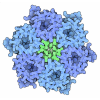
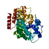
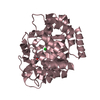
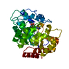
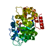
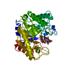
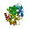
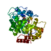
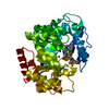
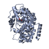
| Title | Crystal structure of adenosine deaminase ligated with EHNA | ||||||
 Components Components | Adenosine deaminase | ||||||
 Keywords Keywords | HYDROLASE / beta barrel / protein-inhibitor complex / Acetylation / Nucleotide metabolism / Pharmaceutical / Polymorphism | ||||||
| Function / homology |  Function and homology information Function and homology information2'-deoxyadenosine deaminase activity / negative regulation of adenosine receptor signaling pathway / cytoplasmic vesicle lumen / purine ribonucleoside monophosphate biosynthetic process / inosine biosynthetic process / adenosine deaminase / hypoxanthine salvage / adenosine catabolic process / adenosine deaminase activity / nucleotide metabolic process ...2'-deoxyadenosine deaminase activity / negative regulation of adenosine receptor signaling pathway / cytoplasmic vesicle lumen / purine ribonucleoside monophosphate biosynthetic process / inosine biosynthetic process / adenosine deaminase / hypoxanthine salvage / adenosine catabolic process / adenosine deaminase activity / nucleotide metabolic process / anchoring junction / T cell activation / lysosome / cell adhesion / external side of plasma membrane / zinc ion binding / cytosol Similarity search - Function | ||||||
| Biological species |  | ||||||
| Method |  X-RAY DIFFRACTION / X-RAY DIFFRACTION /  FOURIER SYNTHESIS / Resolution: 2.52 Å FOURIER SYNTHESIS / Resolution: 2.52 Å | ||||||
 Authors Authors | Kinoshita, T. | ||||||
 Citation Citation |  Journal: Biochem.Biophys.Res.Commun. / Year: 2008 Journal: Biochem.Biophys.Res.Commun. / Year: 2008Title: Conformational change of adenosine deaminase during ligand-exchange in a crystal Authors: Kinoshita, T. / Tada, T. / Nakanishi, I. | ||||||
| History |
|
- Structure visualization
| Structure viewer | Molecule:  Molmil Molmil Jmol/JSmol Jmol/JSmol |
|---|
- Downloads & links
- Download
| PDBx/mmCIF format |  2z7g.cif.gz 2z7g.cif.gz | 85.9 KB | Display |  PDBx/mmCIF format PDBx/mmCIF format |
|---|---|---|---|---|
| PDB format |  pdb2z7g.ent.gz pdb2z7g.ent.gz | 63.3 KB | Display |  PDB format PDB format |
| PDBx/mmJSON format |  2z7g.json.gz 2z7g.json.gz | Tree view |  PDBx/mmJSON format PDBx/mmJSON format | |
| Others |  Other downloads Other downloads |
-Validation report
| Arichive directory |  https://data.pdbj.org/pub/pdb/validation_reports/z7/2z7g https://data.pdbj.org/pub/pdb/validation_reports/z7/2z7g ftp://data.pdbj.org/pub/pdb/validation_reports/z7/2z7g ftp://data.pdbj.org/pub/pdb/validation_reports/z7/2z7g | HTTPS FTP |
|---|
-Related structure data
| Related structure data |  1krmS S: Starting model for refinement |
|---|---|
| Similar structure data |
- Links
Links
- Assembly
| Deposited unit | 
| ||||||||
|---|---|---|---|---|---|---|---|---|---|
| 1 |
| ||||||||
| Unit cell |
|
- Components
| #1: Protein | Mass: 40341.672 Da / Num. of mol.: 1 / Source method: isolated from a natural source / Source: (natural)  |
|---|---|
| #2: Chemical | ChemComp-ZN / |
| #3: Chemical | ChemComp-EH9 / ( |
| #4: Water | ChemComp-HOH / |
| Sequence details | THE SEQUENCE IS BASED ON REFERENCE 2 IN THE DATABASE, ADA_BOVIN. GLN 196, THR 243, AND ARG 349 ARE ...THE SEQUENCE IS BASED ON REFERENCE 2 IN THE DATABASE, ADA_BOVIN. GLN 196, THR 243, AND ARG 349 ARE VARIANTS OF ADA_BOVIN. |
-Experimental details
-Experiment
| Experiment | Method:  X-RAY DIFFRACTION / Number of used crystals: 1 X-RAY DIFFRACTION / Number of used crystals: 1 |
|---|
- Sample preparation
| Crystal | Density Matthews: 2.5 Å3/Da / Density % sol: 50.77 % |
|---|---|
| Crystal grow | Temperature: 293 K / Method: vapor diffusion, sitting drop / pH: 6 Details: 2M Ammonium sulfate, 2% MPD, 0.1M MES buffer, pH 6.0, VAPOR DIFFUSION, SITTING DROP, temperature 293K |
-Data collection
| Diffraction | Mean temperature: 100 K |
|---|---|
| Diffraction source | Source:  ROTATING ANODE / Type: RIGAKU ULTRAX 18 / Wavelength: 1.5418 Å ROTATING ANODE / Type: RIGAKU ULTRAX 18 / Wavelength: 1.5418 Å |
| Detector | Type: RIGAKU RAXIS IV++ / Detector: IMAGE PLATE / Date: Jul 31, 2007 / Details: mirrors |
| Radiation | Monochromator: graphite / Protocol: SINGLE WAVELENGTH / Monochromatic (M) / Laue (L): M / Scattering type: x-ray |
| Radiation wavelength | Wavelength: 1.5418 Å / Relative weight: 1 |
| Reflection | Resolution: 2.52→67.03 Å / Num. all: 14440 / Num. obs: 13675 / % possible obs: 94.7 % / Observed criterion σ(I): 0 / Rmerge(I) obs: 0.062 / Net I/σ(I): 13.4 |
| Reflection shell | Resolution: 2.52→2.75 Å / Rmerge(I) obs: 0.167 / Mean I/σ(I) obs: 3.7 / % possible all: 94.5 |
- Processing
| Software |
| ||||||||||||||||||||||||||||||||||||||||||||||||||||||||||||
|---|---|---|---|---|---|---|---|---|---|---|---|---|---|---|---|---|---|---|---|---|---|---|---|---|---|---|---|---|---|---|---|---|---|---|---|---|---|---|---|---|---|---|---|---|---|---|---|---|---|---|---|---|---|---|---|---|---|---|---|---|---|
| Refinement | Method to determine structure:  FOURIER SYNTHESIS FOURIER SYNTHESISStarting model: PDB entry 1KRM Resolution: 2.52→67.03 Å / Isotropic thermal model: isotropic / Cross valid method: THROUGHOUT / σ(F): 0 / σ(I): 0 / Stereochemistry target values: Engh & Huber
| ||||||||||||||||||||||||||||||||||||||||||||||||||||||||||||
| Refinement step | Cycle: LAST / Resolution: 2.52→67.03 Å
| ||||||||||||||||||||||||||||||||||||||||||||||||||||||||||||
| Refine LS restraints |
| ||||||||||||||||||||||||||||||||||||||||||||||||||||||||||||
| LS refinement shell | Resolution: 2.52→2.57 Å / Rfactor Rfree: 0.331 / Rfactor Rwork: 0.251 |
 Movie
Movie Controller



 PDBj
PDBj
